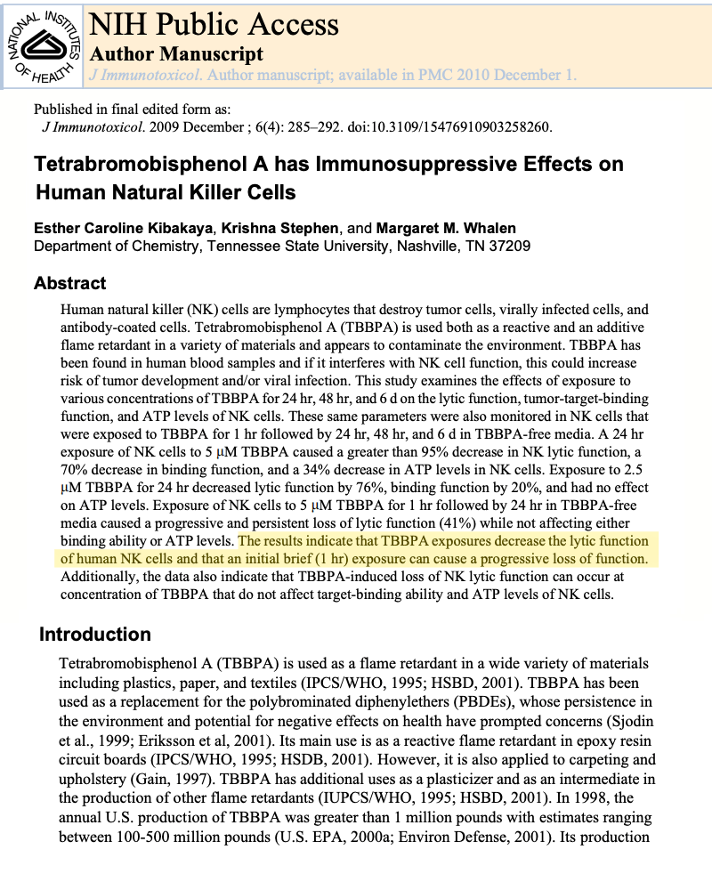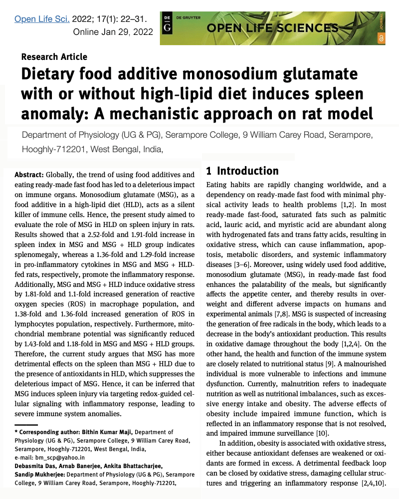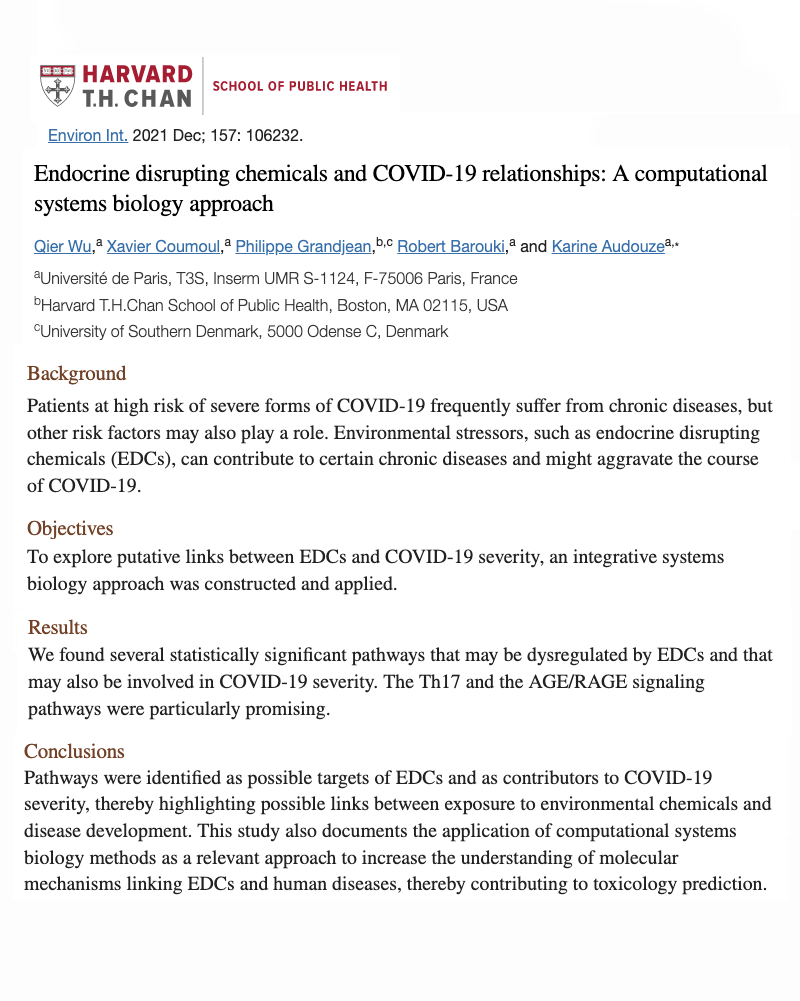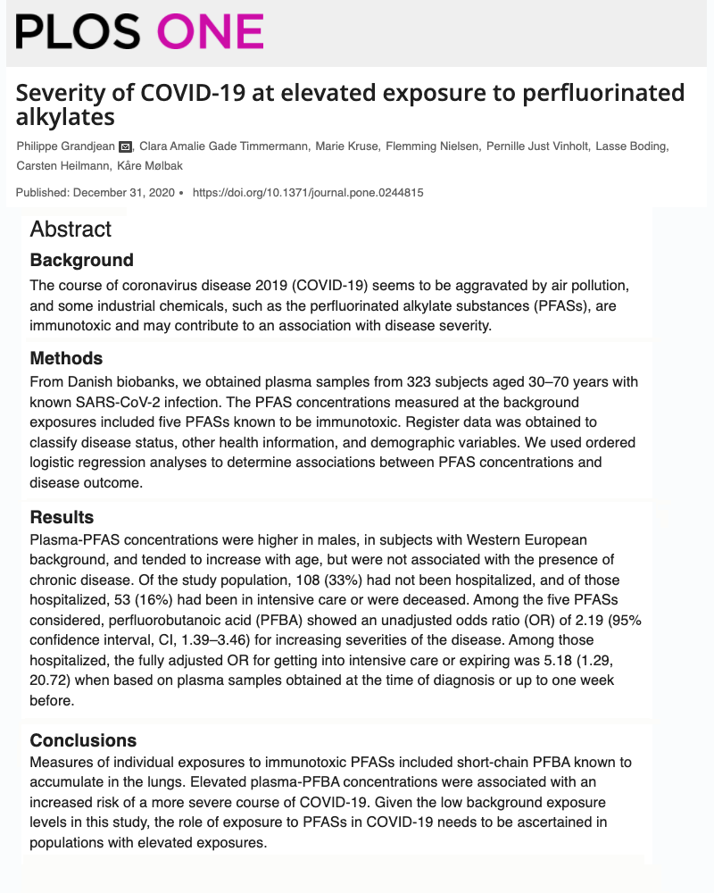
Review of the Medical Literature
INTRODUCTION: Immune system differences are now well documented when comparing those who develop symptoms from COVID-19 to those who show no symptoms (asymptomatic). In order to reverse the increasing trends of viral infections being observed in society today, it is important to not only treat symptoms, but also investigate - Why these immune differences are occurring more frequently than in past years? This website summarizes over 50 peer-reviewed scientific journals that address this question directly, giving insight into environmental factors that significantly weaken immune system function, which in turn increases risk of infection and severity.
Read more for additional background information or scroll below for journal summaries
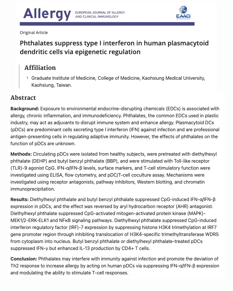
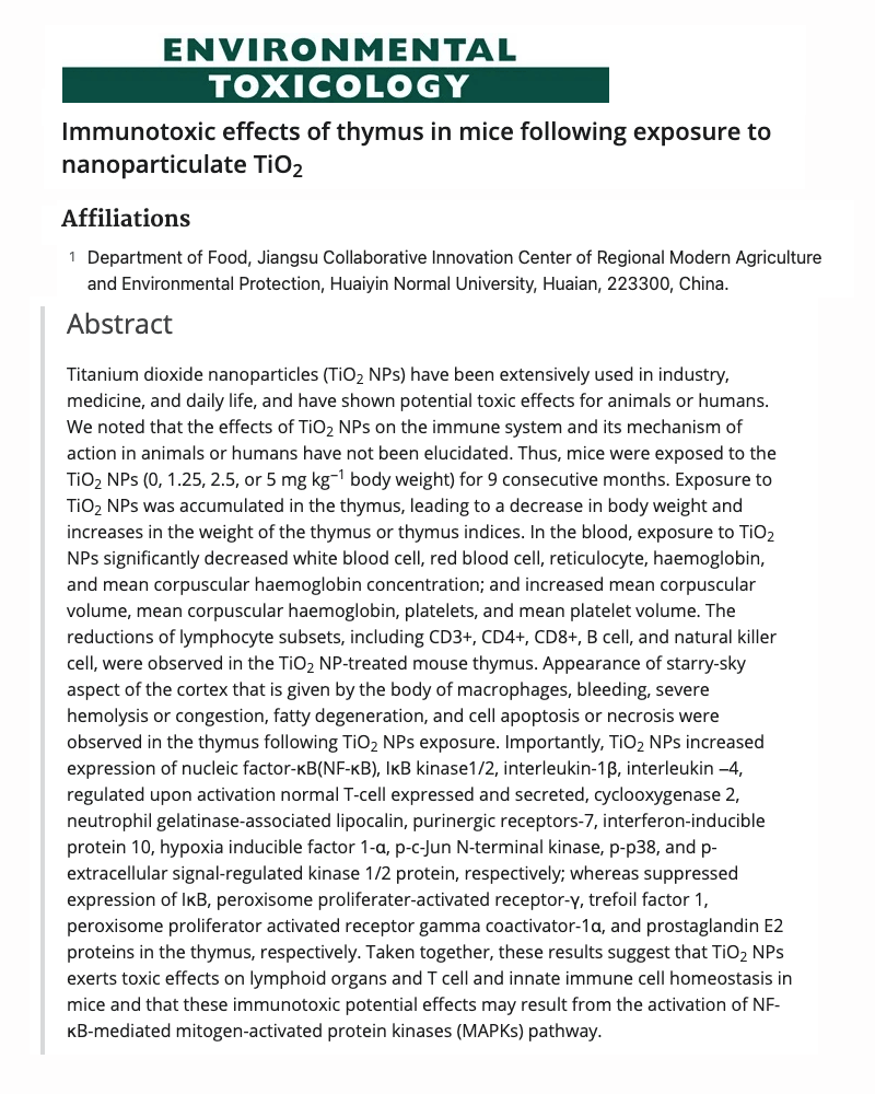
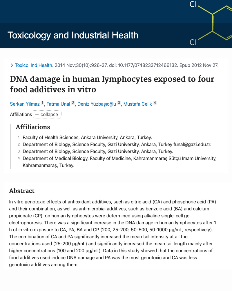
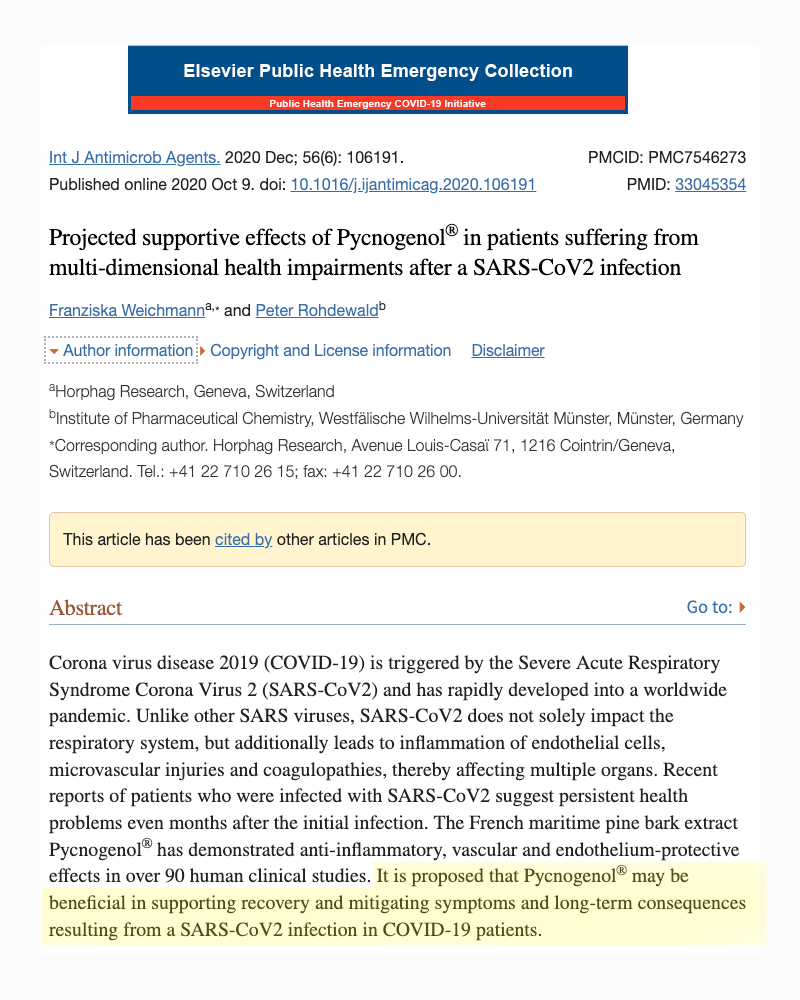
All studies below are peer-reviewed from the National Library of Medicine database (hence the NLM after Corona in our URL). Each post includes a detailed summary of the topic as well as the journal name - research centers involved - a screenshot of the first page of the article abstract and a link to the publicaton in the original journal. A checkbox is located below each post allowing visitors to create their own PDF booklet of the first page "Abstracts" of all articles of interest. Even though the dozens of studies below add great insight into why some develop Covid infection easily and others do not - the conventional television media has chosen not to address these studies. This is a severe disservice to public health as only upon understanding factors that weaken the human immune system, can we then find a way to reverse COVID-19 and other viral infections that are certainly waiting around the corner.
Over 50 studies have now been published documenting air pollution as one of the, if not the largest contributor to the rise in COVID-19. This occurs as the air pollutants nitrogen dioxide and fine particulate matter are inhaled and damage delicate lung tissue. This increases expression of ACE2 receptors, which serve to heal damaged cells but also creates a "doorway" for how the Coronavirus can enter body cells. Air pollultion comes from fossil-fuel power generation (i.e. coal), municipal trash incineration and vehicle traffic, with gasoline and diesel powered vehicles being responsible for an estimated 52% of the problem.
Mucus contains powerful virus killing compounds - but damaged by air pollution
The mucus lining your nose and throat contains a powerful virus killing compound called surfactant protein. These are so effective (and important), they can eliminate viruses long before taking-up residence in your cells. The bad news - inhaling fine particulate matter from vehicle exhaust and local power generation greatly weakens its virus killing ability.Most people have never heard of surfactant proteins, but they are now being shown to be our critical first defense in stopping viral infections such as COVID-19.
While 90% of our lung cells are involved in taking oxygen from the air and putting it into our blood, we have another 10% of lung cells that are greatly underapprciated. These are called Type-II lung cells and are not involved in oxygen exchange. Instead, their role is to make a mucus compound known as surfactant proteins. This keeps our oxygen giving alveoli lung cells moist so they can contract and expand easily during breathing.
We have four types of surfactant proteins made by our type II lung cells which includes SP-A, SP-B, SP-C and SP-D. The one gaining the most attention with COVID-19 is SP-D.
When looking at SP-D closely, researchers found it has the uncanny ability to bind onto the SARS Coronavirus Spike protein, thereby functioning as a natural antibody and literally blocking infection. It also has the ability to kill the virus directly as well as slow viral growth. So basically, three powerful virus stopping abilities all rolled into one molecule.
AIR POLLUTION DAMAGES SURFACTANT PROTEIN
The problem is, when fossil fuels like gasoline, diesel or coal burn, they create a type of fine particle air pollution called PM2.5. The particle itself is 1/30th the width of a human hair. Once inhaled, the chemical poisons hitching a ride on PM2.5 diminish the ability of type-II lung cells to make surfactant protein. AKA, greatly increased risk of viral infection.
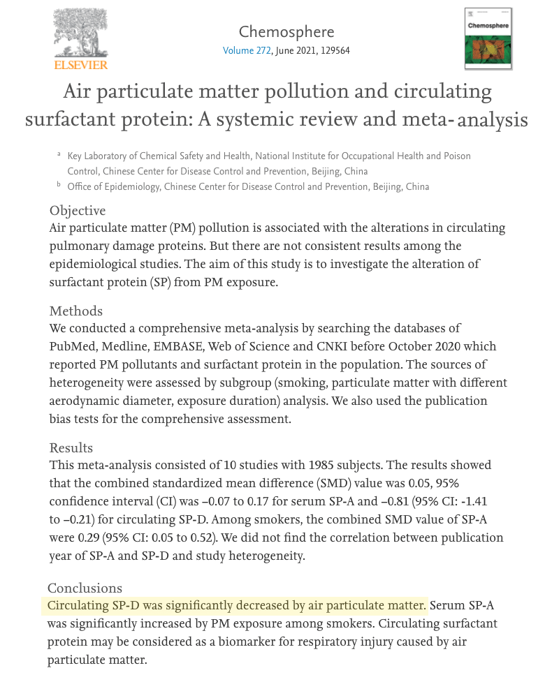
Researchers in this study reviewed 10 separate studies investigating surfactant protein production and damage from air pollution.
Below are relevant quotes from the study's authors beginning on page 6. The letters "PM" represent the term "Particulate Matter and SP-D is the abbreviation for surfactant protein type D.
[BEGIN QUOTES]
1. PM is associated with respiratory damage and even lung cancer incidence (Kelly and Fussell, 2015; Xing et al., 2019).
2. ...experiments have shown that PM2.5 components induced mitochondrial oxidative damage in lung cells and activated DNA damage responses in lymphocytes (Bhargava et al., 2018; Pardo et al., 2019).
3. Exposure to PM has been found to impair the expression of surfactant protein (Silveyra and Floros, 2012).
4. Freberg (Freberg et al., 2016) reported in a long-term PM exposure of 45 Norwegians, SP-D was significantly lower during the exposed days as compared with the non-exposed days. The decrease of surfactant protein may be the result of alveolar type II cell injury and altered synthesis of surfactant (Gregory et al., 1991).
5. Other studies observed that particles can inhibit the expression of SP-A in human alveolar type II cells (Correll et al., 2018; Dong et al., 2019).
6. We found a significant correlation between PM and circulating SP-D in the subgroup analysis stratified by different diameter of particulate matter. However, only PM2.5 was associated with the reduction of circulating SP-D, while PM10 was not.
In their concluding sentence, researchers stated the following:
"Through the subgroup analysis of smoking, aerodynamic diameter and exposure duration of particulate matter, we found that circulating SP-D was significantly decreased by air particulate pollution."
Again, to reiterate, since the lung surfactant protein SP-D is highly effective in stopping viral infection through a multitude of routes, the reduction of lung SP-D production from fine particulate matter from vehicles, power generation, incineration and even backyard trash burning, could be expected to increase COVID-19 infection and severity.
Additional articles on the detailed function of surfactant proteins can be seen in this 2012 review article in Frontiers of Immunology.
https://www.frontiersin.org/articles/10.3389/fimmu.2012.00131/full
Chemosphere
Vol.272:129564, Jun 2021
Chinese Center for Disease Control
and Prevention, Beijing, China.
View Journal HERE
view PDF
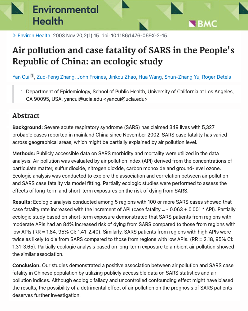
Air pollution increases coronavirus infection by 84%
People living in areas of moderate air pollution had nearly twice the risk of dying from COVID infection as people living in areas of "low" air pollution. Chemicals studied included those typically associated with power generation and vehicle exhaust emission.The School of Public Health at University of California conducted an analysis of SARS coronavirus infections in China. Results showed infection rates were 84% higher in areas with moderate air pollution compared to those living in araas of "low" air pollution. Deaths increased to 118% when comparing high pollution areas with low pollution aeas.
This study adds to the rapidly growing list of scientific research showing background levels of air pollution from fossil-fuel use can increase ACE2 viral receptors and also weaken immune system function, thereby increasing likelihood and severity of viral infections.
VEHICLE EXHAUST CHEMICALS
The air pollution chemicals identified are found in vehicle exhaust and from the burning of coal and other petroleum energy sources. This included particulate matter, sulfur dioxide, nitrogen dioxide, carbon monoxide and ground-level ozone. Again, all of these come from burning of fossil fuels (cars, trucks, coal, etc.)
CROSSING OVER
While fossil fuel burning increases global warming issues that will cause major problems decades from now, this study (and others) show how the burning of fossil fuels is harming each and every one of us at this very moment - continually degrading genes controlling critical immune system cells.
We are now at the point where it appears large numbers of our population are crossing over - entering into a situation where the body can no longer protect from viral infections.
REDUCING IMMUNE SYSTEM
Some of these exhaust chemicals not only can increase ACE2 receptors (giving the virus more locations for binding), but also reduce levels of the immune system hormone interferon inside the lungs and respiratory cells.
Since interferon is one of our first defenses that blocks virus growth - it’s not surprising that viruses increase rapidly among people living in cities with higher background pollution levels.
Environmental Health
Vol.20:2(1), 2003
School of Public Health
University of California
View Journal HERE
view PDF
COVID-19 higher in people with highest 18 year air pollution exposure
First study to show people exposed to higher air pollution the past 18 years had higher COVID infection today. This suggests subtle lung and immune damage occurring from years of "higher" air pollution exposure. Air pollution measured was fine particle matter (PM2.5) which comes mainly from vehicle exhaust, municipal incineration and coal power generation.Background:
COVID-19 is a lung disease, and there is medical evidence that air pollution is one of the external causes of lung diseases. Fine particulate matter is one of the air pollutants that damages pulmonary tissue. The combination of the coronavirus and fine particulate matter air pollution may exacerbate the coronavirus’ effect
on human health.
Research question:
This paper considers whether the long-term concentration of fine particulate matter of different sizes changes the number of detected coronavirus infections and the number of COVID-19 fatalities in Germany.
Study design:
Data from 400 German counties for fine particulate air pollution from 2002 to 2020 are used to measure the long-term impact of air pollution. Kriging interpolation is applied to complement data gaps. With an ecological study, the correlation between average particulate matter air pollution and COVID-19 cases, as well as fatalities, are estimated with OLS regressions. Thereby, socioeconomic and demographic covariates are included.
Main findings:
An increase in the average long-term air pollution of 1 μg/m3 particulate matter PM2.5 is correlated with 199.46 (SD = 29.66) more COVID-19 cases per 100,000 inhabitants in Germany. For PM10 the respective increase is 52.38 (SD = 12.99) more cases per 100,000 inhabitants. The number of COVID-19 deaths were also positively correlated with PM2.5 and PM10 (6.18, SD = 1.44, respectively 2.11, SD = 0.71, additional COVID-19 deaths per 100,000 inhabitants).
Conclusion:
Long-term fine particulate air pollution is suspected as causing higher numbers of COVID-19 cases. Higher long-term air pollution may even increase COVID-19 death rates. We find that the results of the correlation analysis without controls are retained in a regression analysis with controls for relevant confounding factors. Nevertheless, additional epidemiological investigations are required to test the causality of particulate matter air pollution for COVID-19 cases and the severity.
Environmental Research
Vol.204:111948, Mar. 2022
Inst. of Public Economics
University of Muenster
Germany
View Journal HERE
view PDF
Small increase in pollution = Large increase in COVID-19
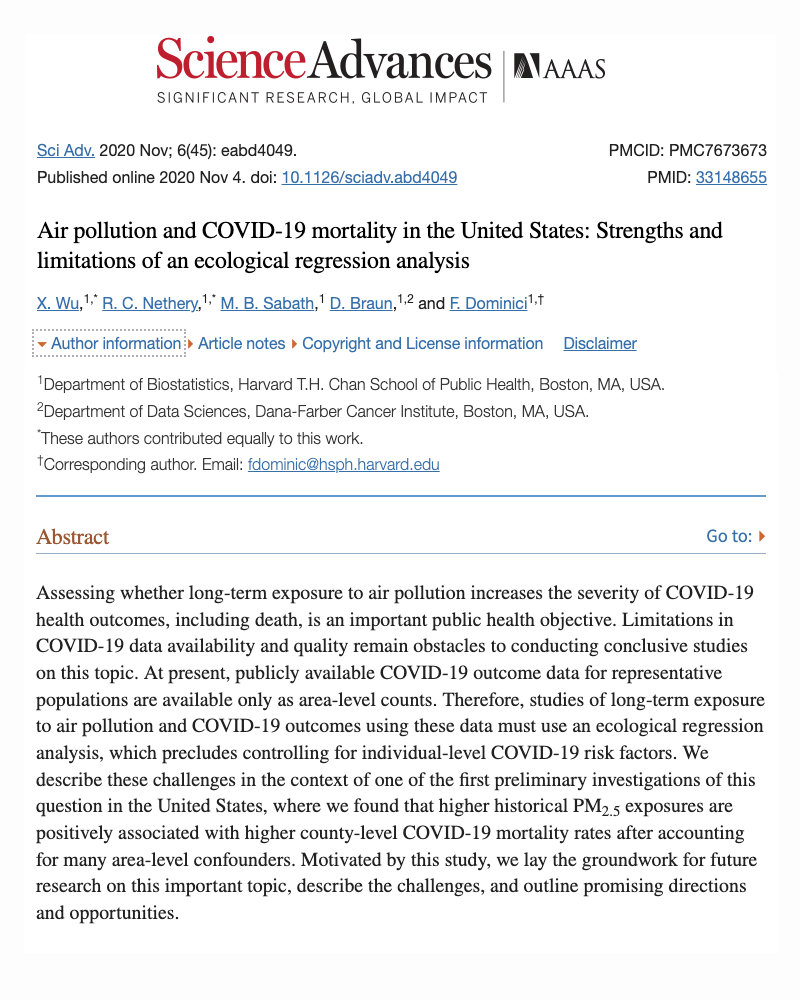
Researchers found people living in areas with only slightly elevated levels of fine particulate matter experienced greatly increased rates of COVID-19. This is believed to occur from air pollution particles causing irritation and inflammation of delicate lung cells, thereby increasing ACE2 viral entry receptors in lungs.Harvard researchers collected air quality data and COVID-19 infection rates from 3,080 counties across the US. Counties with high levels of fine particulate pollution averaged the highest rates of CoronaVirus infections. Fine particle air pollution (also called PM2.5) is only a fraction the width of a human hair. In fact, it takes about 30 PM2.5 particles side by side to equal the width of a human hair. PM2.5 particles are generated largely from gas and diesel combustion from cars and trucks, refineries, municipal incineration, electric power plants and even backyard trash burning.
LENGTH OF TIME EXPOSED IS KEY
The study found that someone who lives for decades in a county with high levels of fine particulate pollution at 13 micrograms per cubic meter is 11% more likely to die from COVID-19 than someone who lives in a region with just 12 micrograms per cubic meter.
The researchers concluded by stating:
"We found that an increase of 1 μg/m3 (1 microgram per cubic meter) in the long-term average PM2.5 is associated with a statistically significant 11% increase in the county’s COVID-19 mortality rate.... The study results underscore the importance of continuing to enforce existing air pollution regulations to protect human health both during and after the COVID-19 crisis."
Science Advances
Vol.6(45),Nov.4,2020
Harvard School of Public Health
View Journal HERE
view PDF
Satellite maps link air pollution to higher Covid infection
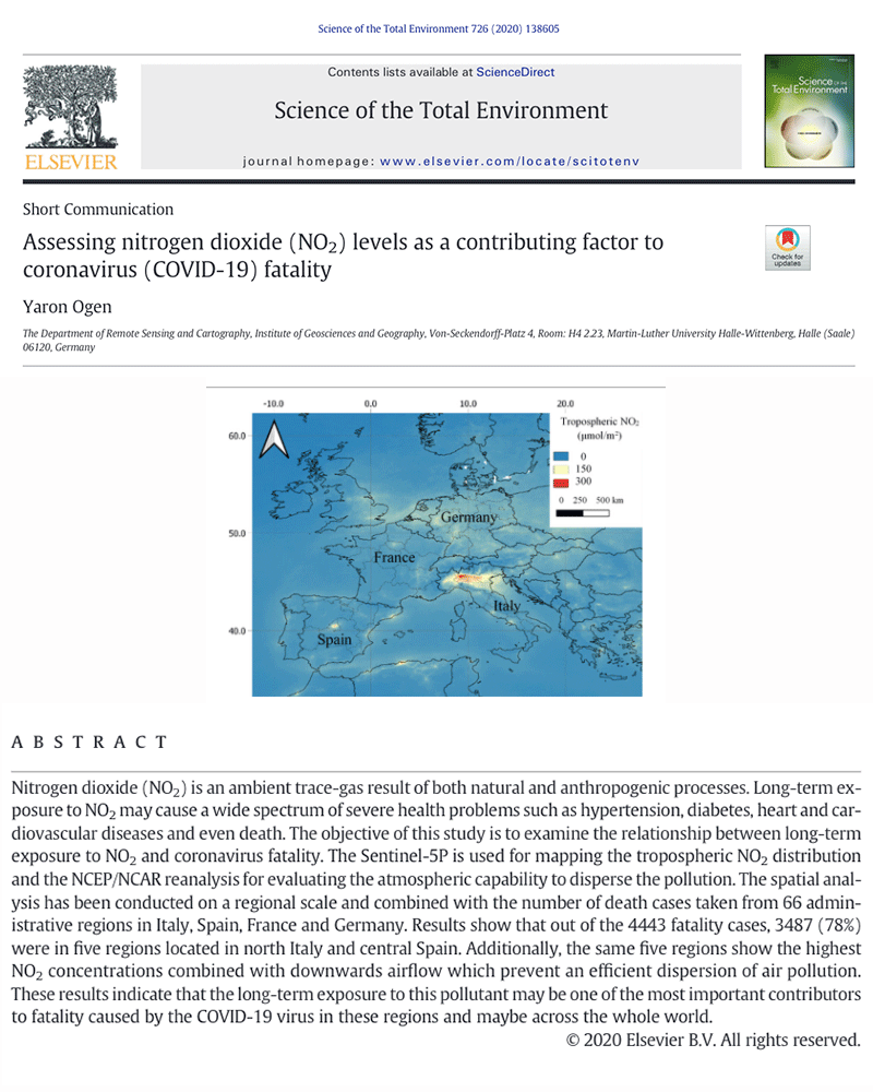
Using satellite mapping technology, higher rates of COVID-19 infection were found to occur in locations with the highest levels of air pollution. Coming mainly from vehicle exhaust and power generation, nitrogen dioxide causes inflammation of the lungs, resulting in higher ACE2 coronavirus docking points.Nitrogen dioxide is a gas formed from the burning of fossil-fuels. Automobiles and trucks emit high levels of nitrogen dioxide and generate 52% of global nitrogen dioxide emissions. This creates highly elevated exposure to those living near busy road-ways or to those who spend extended time driving vehicles (truck drivers - taxis etc).
Long-term exposure to nitrogen dioxide is known to cause inflammation of the lungs and has been linked to severe health problems including high blood pressure, diabetes, heart and cardiovascular diseases and even death (which are also risk factors for Covid-19).
A satellite, called the Sentinel-5P, was launched in 2017 and used for mapping the lower atmosphere for levels of nitrogen dioxide. The nitrogen-dioxide data was combined with the number of COVID deaths taken from 66 administrative regions in Italy, Spain, France and Germany.
Results showed that out of the 4,443 fatality cases - 3,487 (78%) were in five regions located in north Italy and central Spain. Additionally, the same five regions showed the highest nitrogen dioxide concentrations, combined with downwards airflow which prevent an efficient dispersion of air pollution.
The authors concluded by stating,
These results indicate that long-term exposure to this pollutant may be one of the most important contributors to fatality caused by the COVID-19 virus in these regions and maybe across the whole world.
Science of Total Environ.
Vol.726, July 15, 2020
Dept. R Sens. Cartography
Martin-Luther University
Germany
View Journal HERE
view PDF
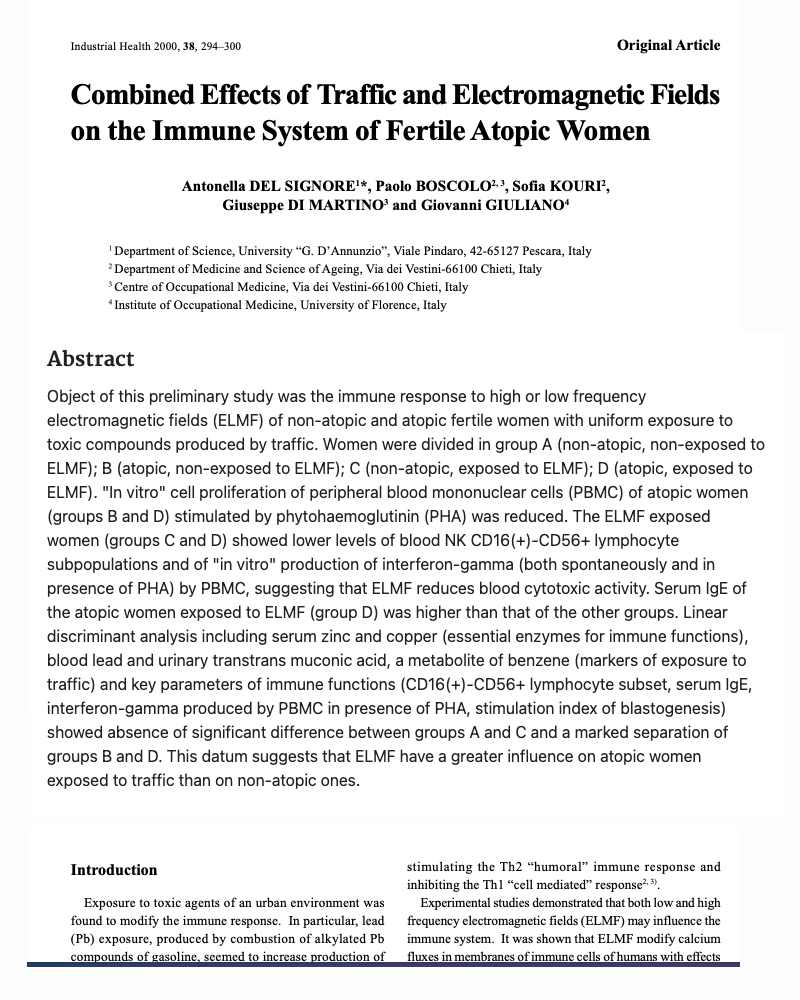
Car exhaust and electromagnetic fields weaken immune system
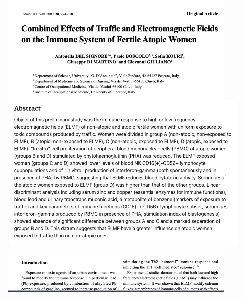
Using women with "allergies" as the test subjects, researchers found simultaneous exposure to both vehicle exhaust and electromagnetic fields (ELMFs) caused a marked reduction in critical immune defenses. This included lower interferon output and decreases in natural killer cells. Interferon and natural killer cells are essential immune defenses needed for fighting COVID-19.In the "real-world," people are not exposed to harmful situations one at a time. In this study, women with and without allergies were exposed to both ELMFs and vehicle exhaust to determine how single and combination exposures affected their immune system. While most lymphocytes were not affected with the levels studied, it was found that exposure to ELMFs reduced interferon (a natural chemical that enhances immune activity) and decreased natural killer cell function.
In conclusion, the researchers stated,
"This result, which demonstrates a different influence of ELMF on non-atopic (non-allergic) fertile women exposed to the toxic compounds of traffic and the non-atopic (non-allergic) ones, suggests that exposure to toxic compounds may increase the effects of ELMF in the atopic (allergic) subjects more than in those non-atopic. (non-allergic)"
Industrial Health
Vol.38(3):294-300, Jul, 2000
University G.D'Annunzio
Italy
View Journal HERE
view PDF
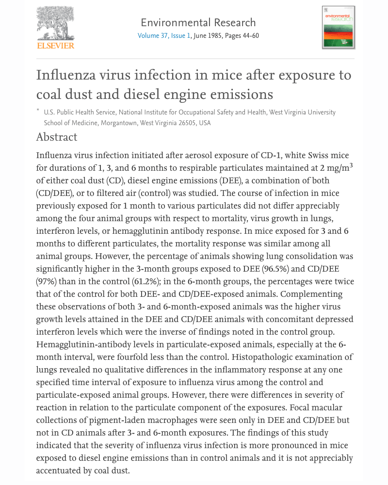
Diesel exhaust increases influenza rates
Mice exposed to diesel exhaust had lower levels of interferon and higher levels of viral growth in lungs. They also produced 4 times less antibodies. This would suggest that vehicle traffic can not only reduce immune defenses, but also reduce vaccine effectiveness as well.White Swiss mice were exposed to diesel engine emissions (DEE), a combination of both (CD/DEE), or to filtered air (control) for durations of 1, 3, and 6 months at 2 mg/m3 of either coal dust (CD). The course of infection in mice previously exposed for 1 month to various particulates did not differ appreciably among the four animal groups with respect to mortality, virus growth in lungs, interferon levels, or hemagglutinin antibody response. In mice exposed for 3 and 6 months to different particulates, the mortality response was similar among all animal groups. However, the percentage of animals showing lung consolidation was significantly higher in the 3-month groups exposed to DEE (96.5%) and CD/DEE (97%) than in the control (61.2%); in the 6-month groups, the percentages were twice that of the control for both DEE- and CD/DEE-exposed animals. Complementing these observations of both 3- and 6-month-exposed animals was the higher virus growth levels attained in the DEE and CD/DEE animals with concomitant depressed interferon levels which were the inverse of findings noted in the control group. Hemagglutinin-antibody levels in particulate-exposed animals, especially at the 6-month interval, were fourfold less than the control. Histopathologic examination of lungs revealed no qualitative differences in the inflammatory response at any one specified time interval of exposure to influenza virus among the control and particulate-exposed animal groups. However, there were differences in severity of reaction in relation to the particulate component of the exposures. Focal macular collections of pigment-laden macrophages were seen only in DEE and CD/DEE but not in CD animals after 3- and 6-month exposures. The findings of this study indicated that the severity of influenza virus infection is more pronounced in mice exposed to diesel engine emissions than in control animals and it is not appreciably accentuated by coal dust.
Environmental Research
Vol.37(3):44-60
West VA Univ School of Med.
View Journal HERE
view PDF
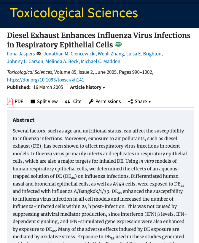
Diesel exhaust increases viral replication inside nasal cavity
A short 2 hour exposure to diesel exhaust greatly increased the rate in which flu viruses were able to multiply inside human nasal and lung cells. Provides important background informatiion for potentially increasing COVID-19 infection.Diesel exhaust contains carbon particles that work like microscopic sponges absorbing a wide variety of toxic chemicals. These are inhaled deep into the lungs while driving on highways or living near high traffic areas.
In a review of other research on the topic, the authors referred to work by Hahon et al.(1985) which showed mice chronically exposed to diesel exhaust for 6 months (and then infected with influenza virus) have decreased ability to produce interferon by 78%. This reduction of interferon led to reduced viral killing and increased viral multiplication. Interferon is essential for blocking virus replication and recruting other immune cells to the area.
Short Diesel Exposure Disarms Defenses In this study from University of North Carolina, researchers exposed live human lung and nasal cell cultures to a liquid solution of diesel exhaust compounds for only two hours. This 2 hour exposure did not reduce interferon like the previous study using a 6 month exposure, but the short exposure did result in a greatly increased growth rate of viruses.
Since interferon was not reduced, the scientists suspected that the large increase in viral growth was at least in part because of a reduction in compounds called Surfactant Protein A and C (called SPA & SPC).
SURFACTANT PROTEINS
These surfactant proteins are important for to learn about because they are literally the first defenders that protect us from viruses. So, if SPA and SPC are doing their job, when you breathe in a virus, the mucus in your airways will catch the virus like a sticky trap, and SPA and SPC then move into action to render the virus harmless. Studies suggest that SPA and SPC can actually wrap themselves around the virus - somewhat analagous to a spider wrapping its prey, and therefore, block the virus from entering the cell.
While much discussion is given to immune system cells and understading their ability to stop an ongoing viral infection, the topic of preventing an infection before it starts is one that needs to be brought into the discussion forefront. Any mechanism that has the ability to cripple either interferon or surfactant proteins, needs to be identified as it is certainly more efficient to prevent a virus from starting than having to deal with several weeks of misery. Additional Studies Linking nitrogen dioxide vehicle exhaust to increased rates of Covid-19 infection. Below are links to 10 of the 15 studies demonstrating that low level air pollution is definitively increasing rates of Covid-19. It's time the media starts talking, before we regret not talking.
Currently, you must copy and paste URL into browser - links will be added shortly.
1. THE NIH STUDY Air pollution and COVID-19 mortality in the United States
https://www.ncbi.nlm.nih.gov/pmc/articles/PMC7277007/
2. Air pollution increases coronavirus deaths in China
https://pubmed.ncbi.nlm.nih.gov/14629774/
3. Role of air pollution in Italy's Covid-19 outbreak.
https://pubmed.ncbi.nlm.nih.gov/32387671/
4. Correlation between air pollution and Covid-19 in California
https://www.ncbi.nlm.nih.gov/pmc/articles/PMC7219392/
5. Short term exposure to air pollution increases Covid-19
https://www.ncbi.nlm.nih.gov/pmc/articles/PMC7159846/
6. COVID-19 spread in northern Italy. The role of pollution?
https://pubmed.ncbi.nlm.nih.gov/32363409/
7. Re-evalutation of pollution's role in viral pandemics
https://www.ncbi.nlm.nih.gov/pmc/articles/PMC7211730/
8. Ozone and nitrogen dioxide linked to Milan, Italy Covid-19
https://www.ncbi.nlm.nih.gov/pmc/articles/PMC7274116/
9. Air pollution and COVID-19 mortality in United States
https://www.ncbi.nlm.nih.gov/pmc/articles/PMC7277007/
10. Air pollution reduction & benefit from COVID-19 lockdown
https://www.ncbi.nlm.nih.gov/pmc/articles/PMC7220178/
Toxicological Sciences
Vol.85(2):990-1002, June, 2005
University of North Carolina,
Chapel Hill
View Journal HERE
view PDF
Truck drivers have unusually high COVID-19 infections
With the previous studies documenting truck and automobile emissions as the biggest risk factor for increasing COVID-19, it shouldn't come as a surprise that nearly 72% of COVID-19 infections were occurring among truck drivers in the African country Uganda. This is not because of person to person contact.Vehicle exhaust has been identified as a major risk factor for increasing COVID-19 infection in over two dozen journals.
The African counry of Uganda has a younger aged population than the United States which provides a unique look into the causes of COVID-19 beyond aging. Below is the abstract discussing higher rates of COVID-19 infection found in truck drivers.
ABSTRACT
Objective
To examine the patterns of COVID-19 transmission in Uganda.
Methods
We reviewed ten weeks of press releases from the Uganda Ministry of Health from the day when the first case was announced, March 22, through May 29, 2020. We obtained the press releases from the MoH website and the Twitter handle (@MinofHealthUG). Data include the number of persons tested and the categories were classified as international arrivals, community members, and long-distance truck drivers.
Results
The first cases were international arrivals from Asia and Europe, and after that, community cases emerged. However, in the middle of April 2020, COVID-19 cases were detected among long-distance truck drivers. By May 29, 2020, 89, 224 persons had been tested; overall, 442 tested positive. Of those that tested positive, the majority, or 317 (71.8%) were truck drivers, 75 (16.9%) were community cases, and 50 (11.3%) were international arrivals. The majority of community cases have been linked to contact with truck drivers.
Conclusions
Truck drivers were the most frequently diagnosed category, and have become a core group for COVID-19 in Uganda. They have generated significant local transmission, which now threatens a full-blown epidemic unless strict controls are put in place.
Inter. J Infectious Diseases
Vol.98:191-193, Sep 2020
Mbarara Univ. Science & Tech.
View Journal HERE
view PDF
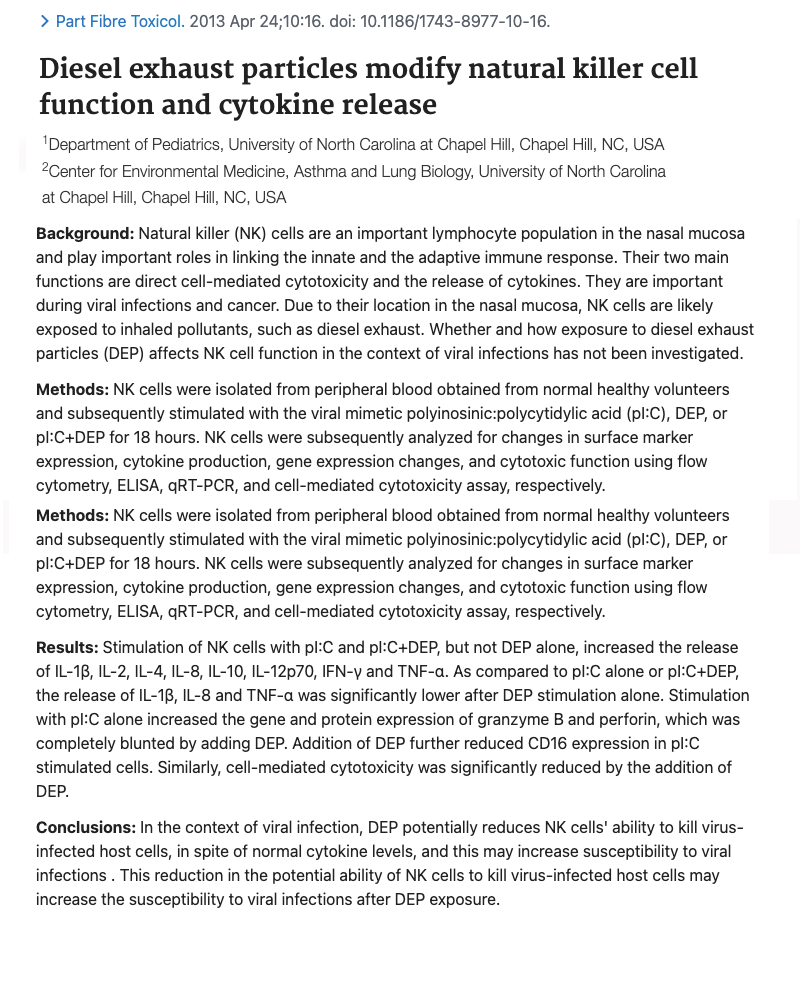
Diesel Exhaust damages Covid-19 defense
Blood donated from 16 University of North Carolina volunteers was exposed to diesel exhaust particles. Testing then demonstrated lower natural killer cell function and decreased ability to remove virus infected cells.For a natural killer cell to "kill" a virus infected cell it must complete three steps:
1. ATTACHMENT
Attach to the cell by identifying it as "stressed" which typically occurs because the cell is missing its MHC marker (which happens after viral infection).
2. OPEN PATHWAY
The NK cell then injects the compound "perforin" which opens a hole in the side of the cell.
3. CELL KILLING
The final step is to inject "granzyme B" into the opening which kills the virus infected cell.
RESULTS
Researchers concluded by stating that diesel exhaust exposure resulted in reduced perforin and granzyme B levels in the NK cell, and hence, lower NK cell function.
Particle Fiber Toxicology
Vol.10:16, April 2013
University of North Carolina
View Journal HERE
view PDF
Studies clearly show those with severe or fatal COVID-19 have distinct immune system abnormalities compared to those with minor infection. While the infection itself can lower numbers and quality of immune cells, it's now been shown that having lower quality immune function prior to infection greatly predisposes someone to severe infection. This can include lower numbers of virus fighting immune cells called natural killer (NK) cells) - improper NK cell function - defective immune cell cytokine communication and high levels of autoimmune antibodies. We'll investigate the growing number of immunology studies in the immune defects section.
Lung surfactant protein protects from COVID-19
10% of our total lung cells are called Type II and manufacture a mucus compound that lubricates our other alveoli lung cells that exhange oxygen during breathing. This mucus also contains substances known as surfactant proteins that have a powerful anti-viral capability and are said to be our first defense in preventing viral infections.Researchers in Germany summarized the current state of knowledge regarding surfactant proteins and how they function as the first defense in stopping viral infections.
Surfactant protein molecules are embedded in the mucus in our lungs, throat and nasal cavity. Once a virus is inhaled and "trapped" in mucus, Surfactant Proteins work to immobilize and destroy the virus.
SURFACTANT PROTEIN BLOCKS VIRAL ENTRY
Another powerful feature of Surfactant Proteins is they have the ability to bind onto the Spike Protein on the SARS-CoronaVirus, thereby functioning as a natural antibody against infection. This study can be seen at -
https://www.frontiersin.org/articles/10.3389/fimmu.2021.641360/full#
WEAKENING OF SURFACTANT PROTEIN PRODUCTION
A number of environmental factors have been identified to reduce the ability of our lung cells to make adequate quantities of Surfactant Protein. The effects from a lack of surfactant protein can be observed in the more advanced stages of COVID-19 viral infection resulting in painful breathing as mucus quantities that moisten type I cells diminish.
Below are key quotes from their article that support the role of Surfactant Proteins in prevention and elimination of COVID-19.
[BEGIN QUOTES]
1. Here it is hypothesized that lowered concentrations or altered composition of pulmonary surfactant is a critical risk factor for COVID-19.
2. Pulmonary surfactant is composed primarily of phospholipids and four surfactant-associated proteins, namely SP-A, SP-B, SP-C, and SP-D. This lipid-protein complex is essential for the biophysical function of the lung. It stabilizes the delicate structure of the mammalian alveoli along with successive compression-expansion respiratory cycles by reducing surface tension at the air-liquid interface (Autilio and Pérez-Gil, 2019).
3. In this regard, most important are SP-A and SP-D, which are members of a family of innate immune proteins termed "COLLECTINS" involved in bacterial and viral clearance (Han and Mallampalli, 2015). These collagen-containing C-type lectins further have ability to opsonize (destroy) pathogens and facilitate their phagocytosis by cells of the innate immune system, such as macrophages and monocytes (Han and Mallampalli, 2015).
4. SP-A (Surfactant Protein A) inhibited influenza A virus infection of lung epithelial cells (Al-Qahtani et al., 2019), while a recombinant fragment of human SP-D was sufficient to act as an entry inhibitor of influenza A virus in vitro (Al-Ahdal et al., 2018).
5. ...it can be concluded that lowering surfactant compounds are causative involved in hypertension presenting a risk factor for developing severe and fatal COVID-19.
6. Some authors speculate that the early administration of natural lung surfactant could improve the pulmonary function to restore pulmonary barrier function in patients with COVID-19 pneumonia, thereby reducing the duration of ventilation therapy and contributing to patients' recovery (Mirastschijski et al., 2020). Indeed, this could be easily done by adding the reconstituted lyophilizate powder into the trachea tube of the ventilated COVID-19 patient.
[END QUOTES]
As environmental factors have been found to weaken the lungs ability to make surfactant proteins (i.e. air pollution and inadequate dietary compounds), this is a cause for concern.
FINAL IMPORTANT NOTE:
When studying Surfactant Protein levels in those with severe COVID-19, patients typically have high levels of Surfactant Protein D (SP-D) in the bloodstream. This occurs because type II lung cells are the first to die from viral expansion. Since SP-D is located inside type II lung cells, when the cell dies, it releases large quantities of SP-D into the blood. While higher lung SP-D levels can be good, having high SP-D in the blood would be considered a bad outcome and evidence of a larger viral lung infection in which lung cells are dying.
Frontiers in Microbiology
Vol. 11:1905, Aug 26, 2020
University Hospital, Aachen, Germany
View Journal HERE
view PDF
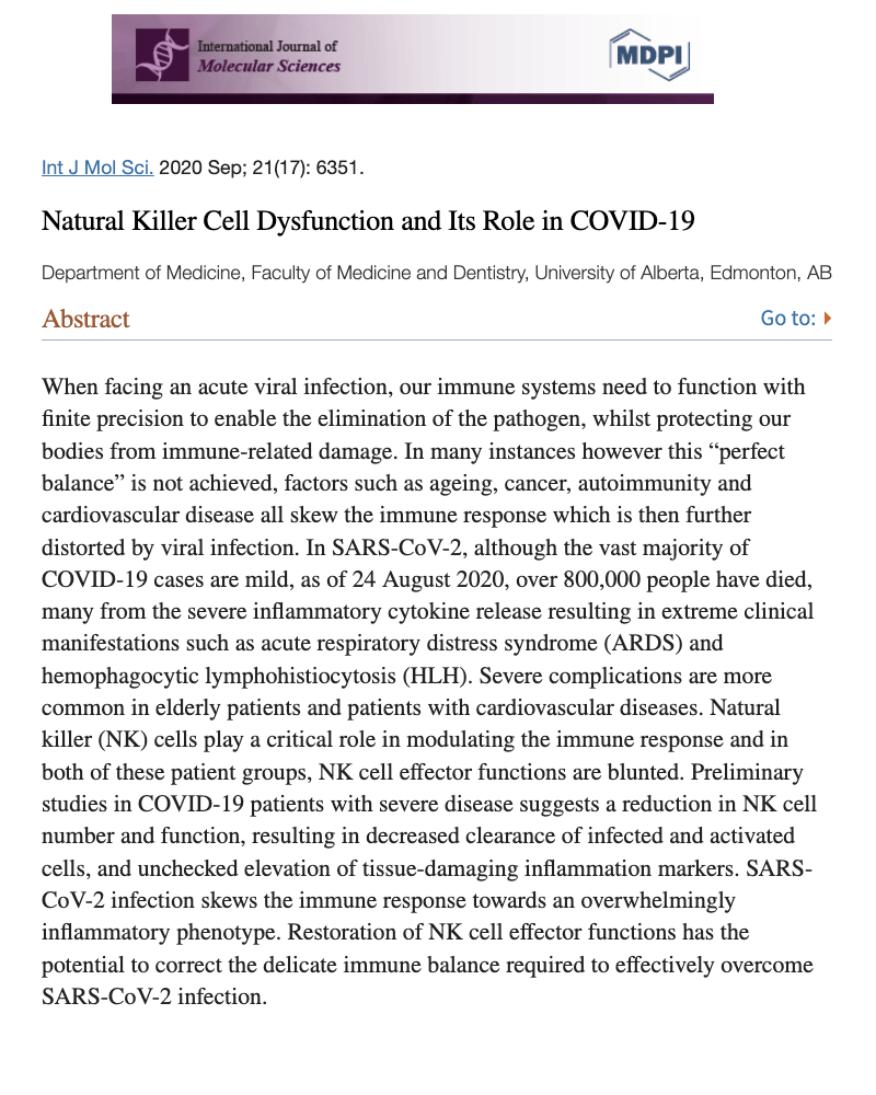
Natural killer cell dysfunction in COVID-19
Natural killer cells are repeatedly noted as the main immune cell that protects from COVID-19 infection. Any disturbance in their function would lead to increased COVID-19 infection and severity. This study found natural killer cell function in COVID-19 patients was "blunted" and suggests ways to improve their function.ABSTRACT
When facing an acute viral infection, our immune systems need to function with finite precision to enable the elimination of the pathogen, whilst protecting our bodies from immune-related damage. In many instances however this “perfect balance” is not achieved, factors such as ageing, cancer, autoimmunity and cardiovascular disease all skew the immune response which is then further distorted by viral infection. In SARS-CoV-2, although the vast majority of COVID-19 cases are mild, as of 24 August 2020, over 800,000 people have died, many from the severe inflammatory cytokine release resulting in extreme clinical manifestations such as acute respiratory distress syndrome (ARDS) and hemophagocytic lymphohistiocytosis (HLH). Severe complications are more common in elderly patients and patients with cardiovascular diseases. Natural killer (NK) cells play a critical role in modulating the immune response and in both of these patient groups, NK cell effector functions are blunted. Preliminary studies in COVID-19 patients with severe disease suggests a reduction in NK cell number and function, resulting in decreased clearance of infected and activated cells, and unchecked elevation of tissue-damaging inflammation markers. SARS-CoV-2 infection skews the immune response towards an overwhelmingly inflammatory phenotype. Restoration of NK cell effector functions has the potential to correct the delicate immune balance required to effectively overcome SARS-CoV-2 infection.
Researchers then go on to suggest factors that can improve natural killer function - including exercise.
Inter. J Molecular Science
Vol.21(17):6351, Sep 2020
Dept of Medicine
University of Alberta, Can
View Journal HERE
view PDF
Immune defects in diabetes increase COVID-19 risk
Severe COVID-19 was more likely to occur in those with type-2 diabetes. Having high blood sugar in diabetes (hyperglycemia) lowers the number and quality of lymphocyte white blood cells needed to eliminate the virus.This study provides an explanation why people with diabetes experience worse outcomes with COVID-19. Apparently, as a person's blood sugar increases, critical immune cells known as lymphocytes decrease. Lymphocytes are approximately 1/3rd of all our white blood cells. Cells affected included helper CD4 T cells, killer CD8 T cells, and natural killer cells. Below is the abstract from the journal:
Abstract
Coronavirus disease 2019 (COVID-19) has been declared a global pandemic. COVID-19 is more severe in people with diabetes. The identification of risk factors for predicting disease severity in COVID-19 patients with type 2 diabetes mellitus (T2DM) is urgently needed.
Methods
Two hundred and thirty-six patients with COVID-19 were enrolled in our study. The patients were divided into 2 groups: COVID-19 patients with or without T2DM. The patients were further divided into four subgroups according to the severity of COVID-19 as follows: Subgroup A included moderate COVID-19 patients without diabetes, subgroup B included severe COVID-19 patients without diabetes, subgroup C included moderate COVID-19 patients with diabetes, and subgroup D included severe COVID-19 patients with diabetes. The clinical features and radiological assessments were collected and analyzed. We tracked the dynamic changes in laboratory parameters and clinical outcomes during the hospitalization period. Multivariate analysis was performed using logistic regression to analyze the risk factors that predict the severity of COVID-19 with T2DM.
Results
Firstly, compared with the nondiabetic group, the COVID-19 with T2DM group had a higher erythrocyte sedimentation rate (ESR) and levels of C-reactive protein (CRP), interleukin 6 (IL-6), tumor necrosis factor alpha (TNF-α), and procalcitonin (PCT) but lower lymphocyte counts and T lymphocyte subsets, including CD3+ T cells, CD8+ T cells, CD4+ T cells, CD16 + CD56 cells, and CD19+ cells. Secondly, compared with group A, group C had higher levels of Fasting blood glucose (FBG), IL-6, TNF-α, and neutrophils but lower lymphocyte, CD3+ T cell, CD8+ T cell, and CD4+ T cell counts. Similarly, group D had higher FBG, IL-6 and TNF-α levels and lower lymphocyte, CD3+ T cell, CD8+ T cell, and CD4+ T cell counts than group B. Thirdly, binary logistic regression analysis showed that HbA1c, IL-6, and lymphocyte count were risk factors for the severity of COVID-19 with T2DM. Importantly, COVID-19 patients with T2DM were more likely to worsen from moderate to severe COVID-19 than nondiabetic patients. Of note, lymphopenia and inflammatory responses remained more severe throughout hospitalization for COVID-19 patients with T2DM.
Conclusion
Our data suggested that COVID-19 patients with T2DM are more likely to develop severe COVID-19 than those without T2DM and that hyperglycemia associated with the lymphopenia and inflammatory responses in COVID-19 patients with T2DM.
ADDITIONAL NOTE: As diabetes has increased over 10-fold since 1960 (going from 1% to over 12% - and now doubling every 12-15 years, this would imply a worsening future for COVID-19 severity.
J Diabetes Complications.
Vol.35(2):107809 Feb 2021
Department of Endocrinology
General Hospital of Central Theater Command
View Journal HERE
view PDF
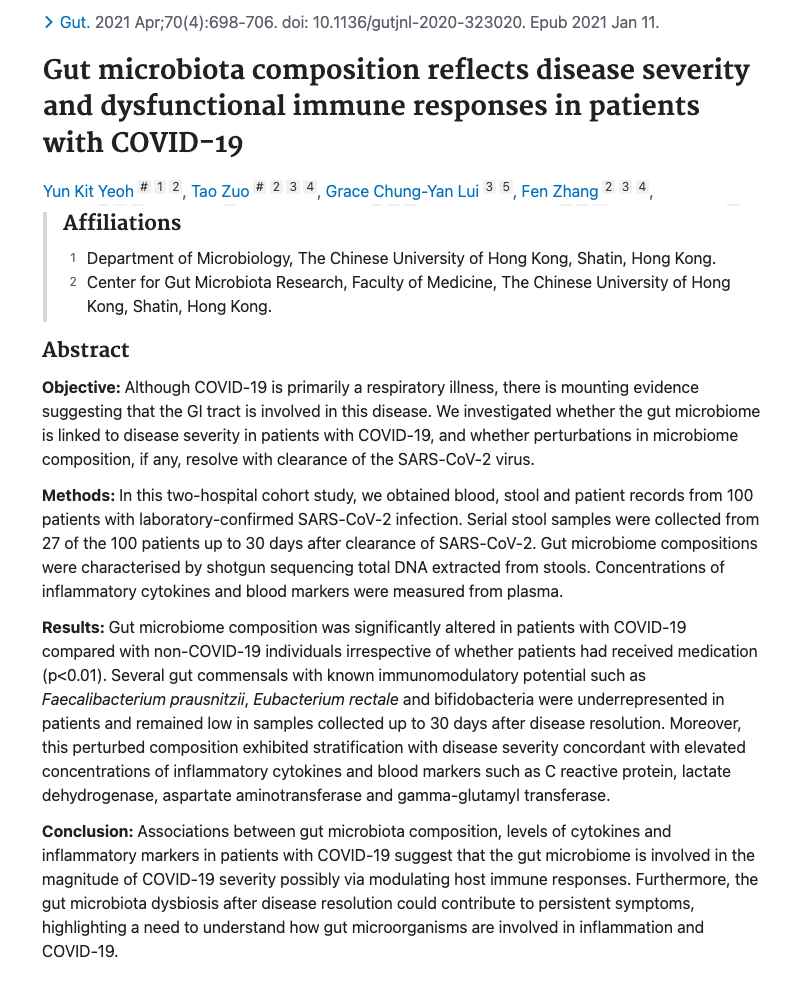
COVID-19 patients have abnormal gut bacteria (microbiota) causing dysfunctional immunity
The trillions of beneficial bacteria in our small and large intestine emit compounds that trigger important immune system functions. These benefical bacteria can be decreased by exposure to various environmental factors in food and air, thereby weakening immune function.Below is the original journal abstract describing the lack of beneificial gut bacteria in those with COVID-10.
ABSTRACT
Objective
Although COVID-19 is primarily a respiratory illness, there is mounting evidence suggesting that the GI tract is involved in this disease. We investigated whether the gut microbiome is linked to disease severity in patients with COVID-19, and whether perturbations in microbiome composition, if any, resolve with clearance of the SARS-CoV-2 virus.
Methods
In this two-hospital cohort study, we obtained blood, stool and patient records from 100 patients with laboratory-confirmed SARS-CoV-2 infection. Serial stool samples were collected from 27 of the 100 patients up to 30 days after clearance of SARS-CoV-2. Gut microbiome compositions were characterised by shotgun sequencing total DNA extracted from stools. Concentrations of inflammatory cytokines and blood markers were measured from plasma.
Results
Gut microbiome composition was significantly altered in patients with COVID-19 compared with non-COVID-19 individuals irrespective of whether patients had received medication (p<0.01). Several gut commensals with known immunomodulatory potential such as Faecalibacterium prausnitzii, Eubacterium rectale and bifidobacteria were underrepresented in patients and remained low in samples collected up to 30 days after disease resolution. Moreover, this perturbed composition exhibited stratification with disease severity concordant with elevated concentrations of inflammatory cytokines and blood markers such as C reactive protein, lactate dehydrogenase, aspartate aminotransferase and gamma-glutamyl transferase.
Conclusion
Associations between gut microbiota composition, levels of cytokines and inflammatory markers in patients with COVID-19 suggest that the gut microbiome is involved in the magnitude of COVID-19 severity possibly via modulating host immune responses. Furthermore, the gut microbiota dysbiosis after disease resolution could contribute to persistent symptoms, highlighting a need to understand how gut microorganisms are involved in inflammation and COVID-19.
Many environmental factors have now been identified (reported previously) that damage microbiota (gut bacteria) function. This includes saturated fat (in red meat) - disinfectants - pesticides - as well as phthalates used in cosmetics and other home products. It is also important to note that defective gut bacteria causes abnormal gut barrier fucntion, thereby allowing viruses into the intestinal tract - that would not have entered if the gut mucus/barrier was intact and working properly.
Gut
Vol.70(4):698-706, Apr 2021
Dept. of Microbiology
Chinese University of Hong Kong
View Journal HERE
view PDF
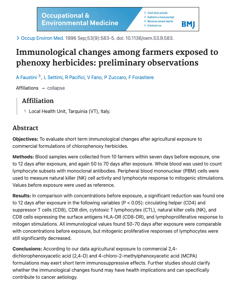
Farmer's show immune dysfunction immediately after pesticide use
Although not directly tied to COVID-19, this study gives clues to COVID-19 outbreaks seen in agriculture areas. Scientists found short-term immune system dysfunction in farmers one to twelve days after applying pesticides on food crops. This included decreased numbers of natural killer cells and CD8 cells - both of which are essential for protecting from COVID-19.What's very interesting about this study is that no immune abnormalities were seen 50-70 days after pesticide exposure. Therefore, other studies looking for immune effects in farmers could easily miss the short-term immune suppression seen here.
Below is the abstract from the farmer/pesticide study. The full PDF gives specific immune counts in a table format for the three time periods of before pesticide spraying - then 1-12 days after and 50-70 days after spraying.
For example, natural killer cells (the main cells needed to prevent viral infection) dropped from nearly 16% of total white count before spraying to 10% one to twelve days after spraying, but then back up to nearly 16% after 50-70 days. Along with natural killer cell numbers decreasing after exposure, their "activity" (how fast they kill virus cells) was less than half as effective after pesticide exposure.
Another cell called CD8-DR dropped by three-fold going from 19% to about 6%. Unlike the CD8 killer cells, the CD8-DR cells work to "suppress" other cells after the virus has been removed. Failure to have enough CD8-DR cells can lead to inflammation and increased autoimmunity.
OBJECTIVES:
To evaluate short term immunological changes after agricultural exposure to commercial formulations of chlorophenoxy herbicides.
METHODS:
Blood samples were collected from 10 farmers within seven days before exposure, one to 12 days after exposure, and again 50 to 70 days after exposure. Whole blood was used to count lymphocyte subsets with monoclonal antibodies. Peripheral blood mononuclear (PBM) cells were used to measure natural killer (NK) cell activity and lymphocyte response to mitogenic stimulations. Values before exposure were used as reference. RESULTS: In comparison with concentrations before exposure, a significant reduction was found one to 12 days after exposure in the following variables (P < 0.05): circulating helper (CD4) and suppressor T cells (CD8), CD8 dim, cytotoxic T lymphocytes (CTL), natural killer cells (NK), and CD8 cells expressing the surface antigens HLA-DR (CD8-DR), and lymphoproliferative response to mitogen stimulations. All immunological values found 50-70 days after exposure were comparable with concentrations before exposure, but mitogenic proliferative responses of lymphocytes were still significantly decreased.
CONCLUSIONS:
According to our data agricultural exposure to commercial 2,4-dichlorophenoxyacetic acid (2,4-D) and 4-chloro-2-methylphenoxyacetic acid (MCPA) formulations may exert short term immunosuppressive effects. Further studies should clarify whether the immunological changes found may have health implications and can specifically contribute to cancer aetiology.
Occupational Environ Med
Vol.53(9):583-5
Sep, 1996
View Journal HERE
view PDF
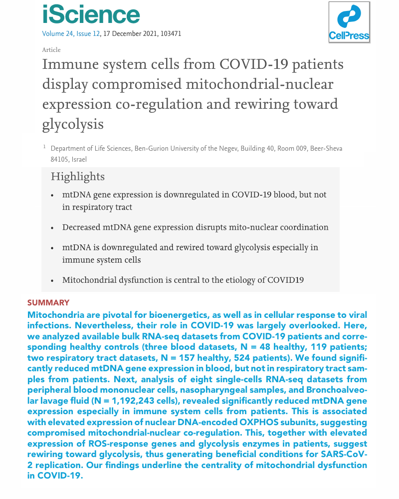
Mitochondria dysfunction in immune cells - Increases COVID-19
Previous studies have shown patients with COVID-19 have abnormal immune function in natural killer cells, T-cells and interferon production. This Israeli study sheds light into why these immune problems occur. Apparently, dysfunctional mitochondria are the source of the problem.Mitchondria are said to be the "energy" of the cell.
While some individuals have only a few hundred mitochondria per cells, other is in very good health have been found to have numbers sometimes approaching 1,000 per cell.
Our immune system cells (ie. natural killer cells, T-cells, etc.) need the "energy" produced by mitochondria to function at optimum level. Any decrease in mitochondria numbers or function would result in cells performing at reduced capacity, thereby weakening immune defenses and increasing risk of viral infections.
Suspecting this might be one explanation for the dysfunctional immune systems seen in basically all hospitalized COVID-19 patients, researchers studied cell mitochondria in blood samples from approximately 800 COVID-19 patients.
When comparing COVID-19 patients with healthy individuals, those with COVID-19 had reduced mitochondrial DNA gene expression in their immune system cells. Since genes control immune cell efficiency, any reduction in gene expression would result in inadequate immune function, thereby increasing risk of infection severity.
Below are additional quotes from the study:
1.Decreased mtDNA gene expression disrupts mito-nuclear coordination.
2.Mitochondrial dysfunction is central to the etiology of COVID19.
As we now have this Israeli study documenting increased damage to mitochondria in COVID-19 patients, this brings up the question of "WHY" mitochondria become dysfunctional in the first place?
We address this topic in the Toxicology section which includes a number of studies showing common endocrine disrupting chemicals (EDCs) cause apoptosis (death) of mitochondria.
Along with EDCs causing mitochondria apoptosis, common EDCs (such as phthalates) were found in one study to suppress genes responsible for mitochondria replication. In other words, we also have documentation showing at least one common household EDC (a specific phthalate) can reduce mitochondria in immune cells, which of course, is highly detrimental to a positive COVID-19 outcome.
iScience
Vol.24:12(17), Dec 2021
Ben-Gurion University of Negev
Israel
View Journal HERE
view PDF
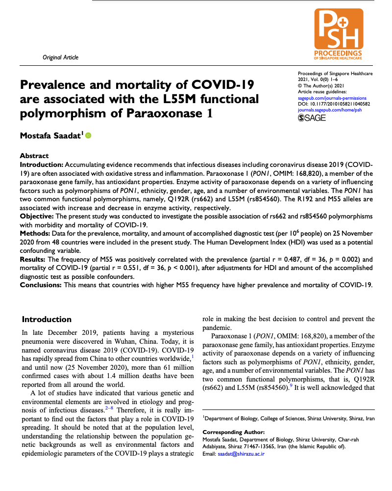
Defects in liver detoxification increases COVID mortality
Not exactly an immune system issue - this study found patients dying of COVID-19 had defects in their liver's ability to make an important detoxification enzyme called Paraoxonase. This would lead to unusually high blood levels of toxic chemicals as well as elevated cell stress (aka too many free-radicals). This in turn results in accelerated damage to cells throughout the body - including the immune system.For you to stay healthy, it is essential that your body can quickly remove the low levels of environmental poisons we are all exposed to that damage our brain or weaken our immune and other defenses.
The biggest player in our toxin removal process is the liver. It accomplishes this through liver enzymes known as cytochrome P450. The CP-450 enzyme low in Covid patients in called paraoxonase. When working properly, it can work as an antioxidant to reduce damage to cells and can also break-apart toxic molecules into a less toxic form. Although not stated in this study, our paraoxonase enzyme can be be weaked by exposure to other toxic chemicals in the environment. This will be discussed later.
Below is the journal abstract which states how those with fatal COVID-19 have mutations in the genese involved in production of the liver enzyme Paroxonase (also known as PON1).
Abstract
Introduction
Accumulating evidence recommends that infectious diseases including coronavirus disease 2019 (COVID-19) are often associated with oxidative stress and inflammation. Paraoxonase 1 (PON1, OMIM: 168,820), a member of the paraoxonase gene family, has antioxidant properties. Enzyme activity of paraoxonase depends on a variety of influencing factors such as polymorphisms of PON1, ethnicity, gender, age, and a number of environmental variables. The PON1 has two common functional polymorphisms, namely, Q192R (rs662) and L55M (rs854560). The R192 and M55 alleles are associated with increase and decrease in enzyme activity, respectively.
Objective
The present study was conducted to investigate the possible association of rs662 and rs854560 polymorphisms with morbidity and mortality of COVID-19.
Methods
Data for the prevalence, mortality, and amount of accomplished diagnostic test (per 106 people) on 25 November 2020 from 48 countries were included in the present study. The Human Development Index (HDI) was used as a potential confounding variable.
Results
The frequency of M55 was positively correlated with the prevalence (partial r = 0.487, df = 36, p = 0.002) and mortality of COVID-19 (partial r = 0.551, df = 36, p < 0.001), after adjustments for HDI and amount of the accomplished diagnostic test as possible confounders.
Conclusions
This means that countries with higher M55 frequency have higher prevalence and mortality of COVID-19.
Proceedings of Singapore Healthcare
September 25, 2021
Deptartment of Biology
Shiraz University, Iran
View Journal HERE
view PDF
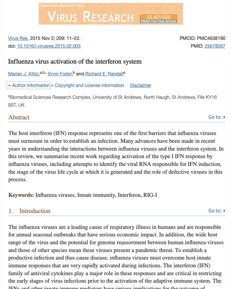
Type 1 Interferon: Understanding the Paul Revere of virus infection
The presence of the immune stimulating hormone interferon is critical if you are to avoid virus infection. It is basically a communication signal between immune cells telling them how to respond once a virus enters your body. As more studies document environmental circumstances that weaken our interferon response - it's time to learn more about this important viral defense.When these soldiers are unable to stop a virus, you have a second line of defense that comes into play, part of that defense is called interferon.
Interferon is not an immune system cell, but simply a compound with a tremendous ability to not only recruit other immune cells to the area, but can also block viruses from entering nearby healthy cells.
However, when these first defenders are weakened and fail in stopping a virus, that's when your remaining immune system army is called in to help.
Once a virus sets up camp on the outside of its chosen respiratory cell, it injects its baby making machinery deep into the cell. At that exact moment, the cell is notified immediately. It sends a flag up to its surface which can literally tell immune system it has been hijacked and that it needs to be destroyed.
Along with this, the cell apparently cares greatly about its neighboring cells. and broadcasts a warning to all cells in the vicinity. This warning is a natural chemical called interferon and has the powerful ability to block other viruses from entering his friends. Unfortunately, scientists are finding a number of situations can reduce the volume of this siren, and therefore, allow for viruses to grow
It was discussed earlier how our lymphocyte white blood cells protect us from basically all virus infections. We discussed how they were found to be critically low in people with fatal COVID-19 outcomes. Another team member, also on the front lines protecting from viral infection, is called Type-1 and Type-2 Interferon (taken from the word interfere).
The studies on this topic suggest that while Type 2 interferon is involved more in removing the virus once infected, Type-1 plays the most important role by preventing the virus from successfully replicating after infecting the first cell in your throat or lung (for Corona) or in the nasal cavity (as with common cold rhinoviruses).
Interferon is not a cell like your lymphocyte white blood cells, but is a powerful chemical messenger that hides below the surface inside apparently all body cells - including cells in your nose, throat and lungs (with the latter two locations being the main growth area for the CoronaVirus.
If a virus makes it past the "sticky" mucus protection covering these cells (another defender and another topic), the virus injects its viral baby-making contents into a cell (and if like a cold virus), 20 minutes later another 100 viral babies come out to carry on the dirty work. However, if the nasal or lung cell is working properly, at the moment the virus enters the cell, it turns on a siren known as type-1 interferon.
This chemical messenger is the Paul Revere of the human immune system and literally warns adjacent cells that the Viruses are Coming - the Viruses are Coming. This causes adjacent cells to go through a biological lock-down sequence that literally prevents entry of any new viral babies that make it out from the first infected cell.
Call in the Cavalry
Not only does type-1 interferon warn cells to lock their doors to block the identified virus attack, but this chemical messenger also calls in the Cavalry to literally attack and destroy any respiratory cells that did succumb to the virus entering the cell. The Calvary in this case, is a group of fighter cells called Natural Killer Cells. (another type of lymphocyte white blood cell). These powerful soldiers have the ability to inject poisons into a virus infected cell to kill it on the spot. This secondary role of interferon was reported in the Journal of Immunology, 135(2):1145-52
Considering the two critical roles of type-1 interferon, it becomes clear that any situation that reduces the ability of our respiratory cells to produce interferon could quickly change a viral infection status from minor to severe.
Virus Research
Vol.209:11-22, Nov. 2015
University of St. Andrews, UK
University of Oxford, UK
View Journal HERE
view PDF
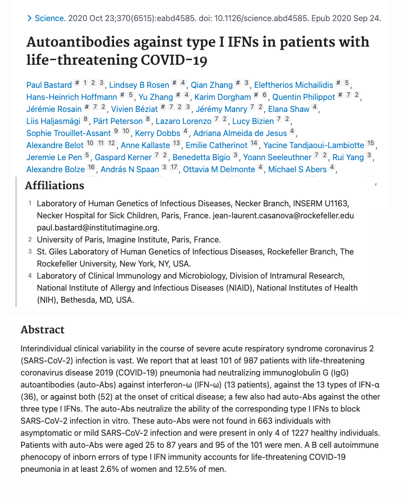
Severe COVID-19 have autoantibodies against type-1 inteferon
Type-1 interferon is a critically important immune system hormone that prevents viral growth in cells and activates natural killer cells to attack the SARS coronavirus. 10% of patients with severe COVID-19 had autoantibodies attacking interferon while mild COVID-19 patients did not have this.The topic of autoantibodies attacking a patient's immune system has drawn immense interest in doctors and scientists world-wide. Over 100 scientists from 73 research centers around the world (including the NIH), participated in this study published in the journal Science in October of 2020.
Results showed over 10% of those with severe COVID-19 had autoantibodies against type-1 interferon while those with mild or asymptomatic COVID-19 had no autoantibodies whatsoever.
Interferon type-1 is the first immune communication needed to stop viral infection. The hormone increases dramatically after the SARS coronavirus breaches and infects a cell. Interferon type-1 then functions to block infection of nearby cells and also calls in natural killer cells to eliminate the virus. Since those with severe COVID-19 also have low natural killer cells, this finding of autoimmunity toward interferon is of major importance.
Below is the complete abstract (see the full-text link for more details.
ABSTRACT
Interindividual clinical variability in the course of severe acute respiratory syndrome coronavirus 2 (SARS-CoV-2) infection is vast. We report that at least 101 of 987 patients with life-threatening coronavirus disease 2019 (COVID-19) pneumonia had neutralizing immunoglobulin G (IgG) autoantibodies (auto-Abs) against interferon-ω (IFN-ω) (13 patients), against the 13 types of IFN-α (36), or against both (52) at the onset of critical disease; a few also had auto-Abs against the other three type I IFNs. The auto-Abs neutralize the ability of the corresponding type I IFNs to block SARS-CoV-2 infection in vitro. These auto-Abs were not found in 663 individuals with asymptomatic or mild SARS-CoV-2 infection and were present in only 4 of 1227 healthy individuals. Patients with auto-Abs were aged 25 to 87 years and 95 of the 101 were men. A B cell autoimmune phenocopy of inborn errors of type I IFN immunity accounts for life-threatening COVID-19 pneumonia in at least 2.6% of women and 12.5% of men.
Science
Vol.370(6515), Oct 2020
72 research centers including:
University of Paris, France
NIH (National Inst of Health)
View Journal HERE
view PDF
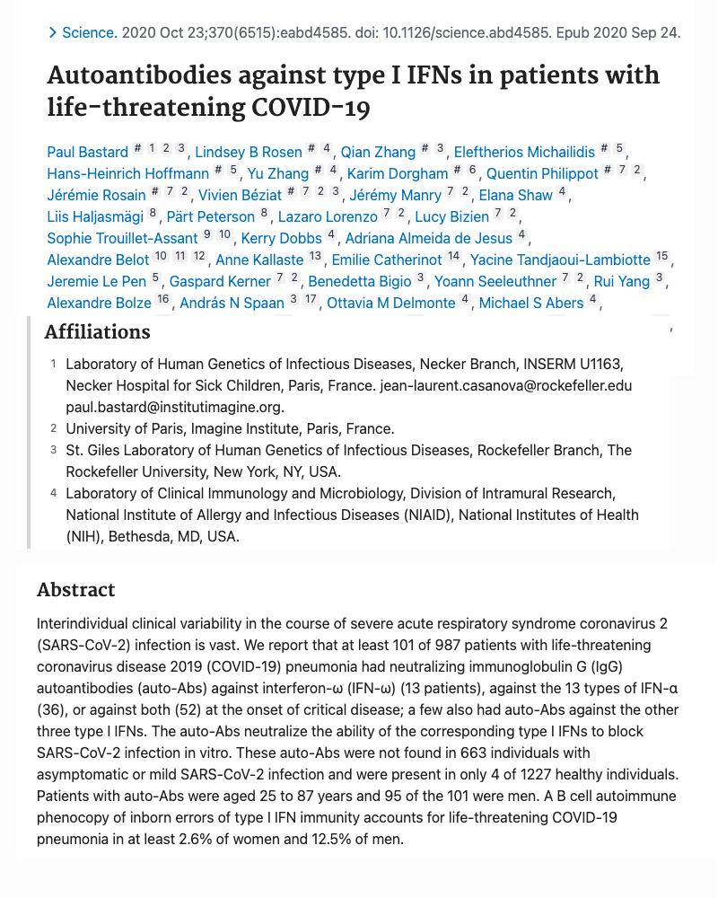
Severe COVID-19 patients have autoantibodies against type-1 inteferon
Interferon type-1 is an essential hormone immune cells use to launch the attack agains the SARS Coronavirus. It can function to block viral entry as well as activate virus killing natural killer cells to increase in number. 10% of patients with severe COVID-19 had a dangerous immune system malfunction resulting in autoantibodies destroying interferon. This was not seen in patients with mild or asymptomatic COVID-19.The topic of autoantibodies attacking a patient's immune system has drawn immense interest among doctors and scientists world-wide. Over 100 scientists from 73 research centers around the world (including the NIH), participated in this study published in the journal Science in October of 2020.
Results showed over 10% of those with severe COVID-19 had autoantibodies against type-1 interferon while those with mild or asymptomatic COVID-19 had no autoantibodies whatsoever.
Interferon type-1 is the first immune communication needed to stop viral infection. The hormone increases dramatically after the SARS coronavirus breaches and infects a cell. Interferon type-1 then functions to block infection of nearby cells and also calls in natural killer cells to eliminate the virus. Since those with severe COVID-19 also have low natural killer cells, this finding of autoimmunity toward interferon is of major importance.
Below is the complete abstract (see the full-text link for more details.
ABSTRACT
Interindividual clinical variability in the course of severe acute respiratory syndrome coronavirus 2 (SARS-CoV-2) infection is vast. We report that at least 101 of 987 patients with life-threatening coronavirus disease 2019 (COVID-19) pneumonia had neutralizing immunoglobulin G (IgG) autoantibodies (auto-Abs) against interferon-ω (IFN-ω) (13 patients), against the 13 types of IFN-α (36), or against both (52) at the onset of critical disease; a few also had auto-Abs against the other three type I IFNs. The auto-Abs neutralize the ability of the corresponding type I IFNs to block SARS-CoV-2 infection in vitro. These auto-Abs were not found in 663 individuals with asymptomatic or mild SARS-CoV-2 infection and were present in only 4 of 1227 healthy individuals. Patients with auto-Abs were aged 25 to 87 years and 95 of the 101 were men. A B cell autoimmune phenocopy of inborn errors of type I IFN immunity accounts for life-threatening COVID-19 pneumonia in at least 2.6% of women and 12.5% of men.
Science
Vol.370(6515), Oct 2020
73 research groups incl.
University of Paris
NIH - National Inst Health
View Journal HERE
view PDF
Severe COVID-19 cases - Low Lymphocytes
Researchers in China found those who died of COVID-19 had extremely low levels of important immune system white blood cells called lymphocytes. Those who survived had more than twice as many. Details on these numbers - how they protect from viruses - how they are made in the body - and why they could be low are also discussed.Lack of discussing readily available information has led to over-exaggerated concerns in many cases and unnecessary hardships for families and the public.
The aim of this section is to clarify what it means to have a "weakened immune system" as it relates to COVID-19. Along with this, it would certainly be prudent to discuss the latest research concerning environmental conditions found to quickly weaken immune system integrity.
LYMPHOCYTES PROTECT FROM VIRUSES
The specifics on what parts of the immune system are weak in patients with serious COVID-19 symptoms was addressed by researchers at the Department of Infectious Disease in Hubei, China.
Doctors found those who died of the CoronaVirus (COVID-19) had dramatically lower numbers of a specific type of immune system white blood cell called a lymphocyte. These are white blood cells have a strong ability to attack and remove all viruses within the body.
Lymphocytes comprise approximiately 30% of all white blood cells in the blood and are "born" in the bone marrow and develop to maturity in the lymmph nodes, tonsils and thymus gland. Most people have lymphocyte levels between 1,100 and 3,100 per cubic millimeter of blood (less than a drop), however, those who died from CoronaVirus infection had far fewer lymphocytes of only 600 per cubic millimeter of blood.
LOW NUMBERS = HIGH RISK
Since lymphocytes are the types of white blood cells that fight a virus infection, having lower numbers before infection is well-known to predispose an individual to a far more serious outcome in any viral infection. In fact, having numbers below 1000 is considered to greatly increase one's risk of serious infection and is medically known as "lymphopenia."
In trying to explain the very low levels of lymphocytes in those who do not survive COVID-19, some have put forth the theory that the CoronaVirus itself infects lymphocytes, however, no research is available to support this, in fact, it is highly unlikely since the receptor that binds the virus to cells (called the ACE2 protein), is not present on lymphocytes (Nature, 426:450-4).
It has also been stated the more likely cause of very low lymphocytes in severe Corona patients, is the recruitment of white blood cells to the site of infection. However, low levels of white blood cells does not happen to all individuals with CoronaVirus infection, therefore, something else must be going on as well.
Other scientists have suggested that the lymphocyte white blood cell production system within the bone marrow and thymus gland becomes "exhausted" - and can no longer keep up with the increased demand for new lymphocytes needed to combat a growing virus.
Whatever the cause of low lymphocytes, it is a fact that nearly 2% to 3% of the adult population already has very low lymphocyte levels below 1000 - even before being infected with the virus.
Therefore, for anyone who develops a COVID infection, it is common sense that going into viral battle with a lymphocyte count of 2000-3000 would be far more advantageous than having a lymphocyte count of 1000 or less. Certainly, a strategy that can be appreciated by anyone with a military background.
Intensive Care Medicine
March 3, 2020
Dept. of Infectious Diseases
Tonji Medical College, China
View Journal HERE
view PDF
Specific lymphocytes low in CoronaVirus patients
Specific types of lymphocyte white blood cells were abnormally low in patients who had severe or fatal reactions to the SARS CornaVirus outbreak in 2002/03. Immune cells identified as low are called Helper T-Cells and Killer T-Cells. The importance of these cells in fighting viruses are discussed..
Helper T-Cells mature in the Thymus gland (hence the letter "T") and give instructions to other immune cells on how best to attack and remove invading viruses. Since Killer T-Cells have the ability to directly kill virus infected cells, low numbers would greatly predispose an indidual to a more severe viral outcome.
WHY LYMPHOCYTES ARE LOW
Although the virus itself can sometimes lead to lower numbers of lymphocytes in someone with an infection (as they are called to the area of infection), the inability of the bone marrow and lymphoid organs to keep up with demand for new cells is a topic of concern.
Inter J of Infectious Disease
Vol.9:323-330, 2005
Capital Univ. of Med Sciences
China
Center Infectious Diseases
Australia
View Journal HERE
view PDF
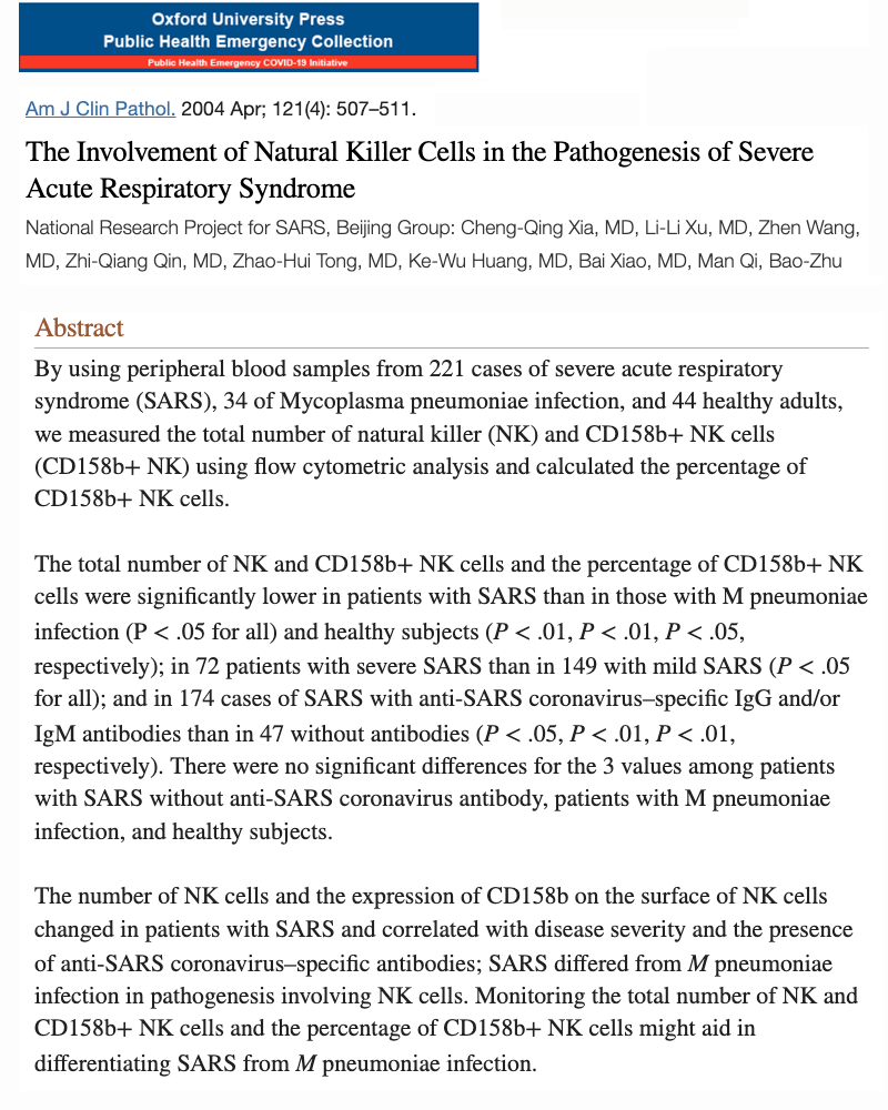
Natural Killer cells low in patients with CoronaVirus (SARS) 2002-2003
Natural Killer cells are being found to be the most important type of lymphocyte white blood cell for eliminating viruses "before" symptomatic infecton appears. People with low numbers of natural killer cells have a far higher risk of viral infections.For example, a type of immune system white blood cell found low in those with the 2002-2003 SARS CoronaVirus is the Natural Killer Cell. These are a specific type of lymphocyte that makes up about 5-10% of all lymphocytes. They are considered to be one of the - if not the first lymphocyte to attack virus infected cells. Therefore, if natural killer cells are working properly (and in sufficient numbers) they would be expected to take control of the any viral situation and kill the first viruses that do infect nasal and/or lung cells.
However, in this study comparing blood samples of 221 patients infected with the CoronaVirus in 2002-2003 with that of 44 healthy adults, it was found that CoronaVirus patients had significantly lower levels of Natural Killer Cells. In other words, this critical part of their first line immune defense was also found to be defective when compared to healthy individuals. This observation should also lead to discussions on factors known to erode proper Natural Killer Cell function.
American J Clinical Pathology
Vol.121(4):507-511, April, 2004
Beijing Chaoyang Hospital, China
National Research Project SARS
View Journal HERE
view PDF
Natural killer cells protect from influenza
Stanford University found students with higher levels of natural killer cells did not contract the flu even after direct nasal injection. This study strongly supports the contention that natural killer cells are the human body's first defense with the ability to prevent viral infections before it can start. This also raises concern as other toxicology studies are finding natural killer cells are far more vulnerable to malfunction following low level exposure to various environmental and chemical circumstances.Researchers at Stanford University have identified a type immune system white blood cell that appears to be responsible for protecting us from the flu.
Dr. Purvesh Khatri, associate professor at Stanford, worked with researchers at Harvard and Duke University to recruit 52 volunteers who generously allowed themselves to be infected with a common influenza virus.
Before inoculating students with the virus (via Q-Tips in the nose), researchers conducted a detailed immune system analysis of all 52 volunteers. After the Q-Tip infection procedure, some students came down with the flu and others did not. The researchers went on to state:
“We found that a type of immune cell called a natural killer cell was consistently low at baseline in individuals who got infected. Those who had a higher proportion of natural killer cells had better immune defenses and fought off illness."
Blood tests of those who came down with the flu showed that less than 10% of their white blood cells were natural killer cells. Students who avoided the flu entirely had 10-13% natural killers. Dr. Khatri went on to state,
It’s a fine line, but the distinction between the groups is quite clear: Everyone who had 10 percent or more natural killer cells stood strong against the infection and showed no symptoms.
While this study was not directed specifically for the COVID-19 virus, it is interesting to note that another study in our report found that COVID-19 patients with a fatal outcome had less natural killer cells than people who recovered.
Genome Mediine
Vol.10:45, June, 2018
Stanford Univ. School of Medicine
Institute for Immunity
View Journal HERE
view PDF
A number of studies have shown cold air increases viral growth in the human body by reducing a critical lung immune defense known as interferon. Interferon increases the effectiveness of various immune cells. Low normal body temperature has also been found to lower body interferon and increase viral growth rate. Higher body temperatures in the fever range have been shown to slow viral growth and increase the number and quality of many immune cells (including natural killer cells).
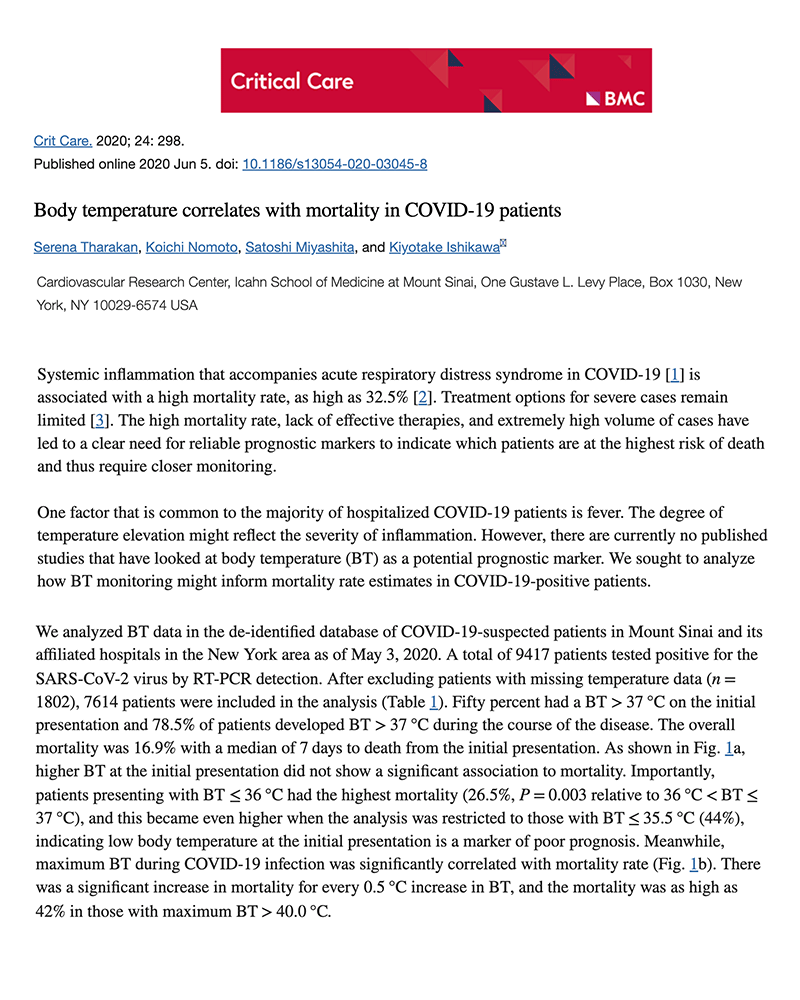
Low body temperature increases COVID deaths
COVID-19 patients with a "low normal" body temperature (below 96.9F or 36C) had nearly twice the risk of dying from COVID-19. Patients with an even lower body temperature (below 96F or 35C) had a nearly 3-times greater risk of dying. This emphasizes the apparent large role body temperature plays in achieving maximum immune system function.The benefits of having a fever in reducing a viral infection has been proven in a number of studies the past few decades. For example, studies of mice given fever lowering aspirin experienced a higher rate of death from viral infections than mice not given aspirin.
This demonstrates the immune benefits of fever and should make us also consider if a "lower normal" body temperature could in some way increase viral infections such as COVID-19. In investigating this subject I could find only one research group who thought to consider this possibility and the results in my view were quite startling.
COMPARING BODY TEMP & COVID DEATHS
Researchers at the Cardiovascular Research Center at Sinai School of Medicine looked at the body temperature records of 7,614 Covid patients at their affiliated Mount Sinai hospitals in the New York area prior to May, 2020.
On the Covid patients' initial check-in to the hospital, it was found that 50% had a body temperature higher than the normal 98.6 degrees F (37C).
During the course of their stay, about 75% of patients developed a fever above that level. Of all 7,614 patients in the study, approximately 17% (about 1 in 6) died, with the median time of death just 7 days after entering the hospital.
LOWER THE TEMP = HIGHER THE DEATHS
Now comes the numbers that were totally unexpected and strongly predicted death. First, it was found that having a high fever-range body temperature above 104F (40C) continuously throughout their infection, was associated with an increased risk of death. However, here's the interesting part - having a body temperature lower than normal resulted in an even higher rate of death.
1. TEMPS BELOW NORMAL
As mentioned, 17% of all COVID patients who entered the hospital in this large study died. But this was just a general statistic with no details regarding body temperature.
However, if the person had a body temperature below or equal to 96.8F or 36C (about 2 degrees below normal F and 1 degree below normal C), it turns out that 26.5% of these COVID patients died (about 1 in 4). But it gets worse.... when looking at Covid patients with a body temp below 96 degrees F (35.5C), their risk of death jumped up to an even higher 44%. This is nearly 3 times higher than the overall 17% death rate.
MY TAKE
This really is fascinating information when you think about it - so nearly half of all Covid patients entering the hospital with a body temperature below 96 degrees F die.
This major clue tells us we better start looking into factors that influence body temperature and how it can be damaged as well as increased.
HOW THE BRAIN CONTROLS BODY TEMP
So, while the outside cortex of our brain controls our personality, speech, and other mental processes, deep inside the lower center of our brain is a pea sized structure called the hypothalamus. Shooting out from this round rascal are nerve fibers that wind up in distant locations on our skin. Here it samples body temperature and relays information back to the hypothalamus, thereby telling it to either raise or lower body temperature.
Taking this altogether, it is certainly reasonable to consider that a low body temperature could occur from a low functioning hypothalamus and/or interrupted or damaged nerve impulses from skin receptors to the hypothalamus.
LOW BODY TEMP HITS HOME
I have a bit of personal experience with low body temperature. For nearly a decade 30 years ago, my normal body temperature was 96.7F (so about 2 degrees below normal). During this time I was catching colds constantly, had zero energy, depression, and my immune white blood count was in the lower 1% of the population.
After talking to some docs in environmental medicine, they recommended i toss the petroleum household based products and eat foods without pesticides and additives. Upon taking their advice, all symptoms and temperature reversed. In fact, even today at age 67, my normal body temperature is 98.3, and that's a lot better than those who die of Covid and quite possibly much higher than those who get symptomatic Covid in the first place and a statistic we need to investigate.
COMMON SENSE TREATMENT FOR COVID-19
Since a far higher number of Covid deaths were shown to occur among those with a low body temperature, this immediately tells us we should investigate potential benefits from raising body temperature manually (in both Covid and Long Covid).
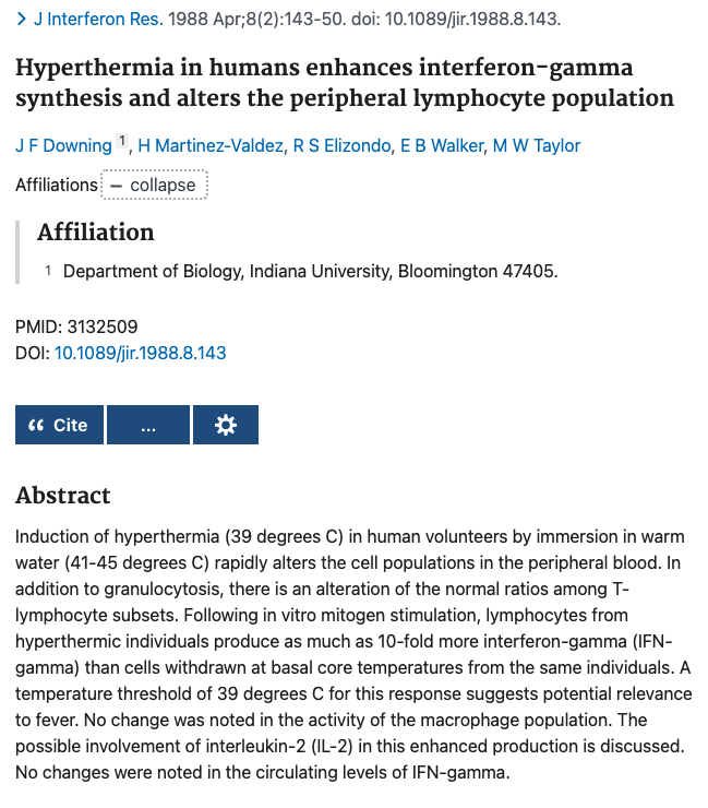
Interestingly, in a separate study of college students, researchers found that putting them in tubs of hot water (and raising their body temps to 101F), resulted in a dramatic improvement in numbers of immune cells called natural killer cells. This high number of good-guy cells lasted over 5 days. Since natural killer cells are powerful killers of flu and Covid virus cells, it certainly seems plausible to give this intervention a try with not only Covid patients, but with Long Covid patients as well.
Organizing and testing hyperthermia treatments in a medical setting with infra-red saunas would be non-invasive and very inexpensive. Unfortunately, it's just this simplicity and low-cost that may prevent this information from reaching those who could benefit the most - and that's a shame...
ATTENTION ALTERNATIVE PRACTITIONERS
However, for alternative medicine practitioners who implement strategies beyond pill ingestion to address health, the link below provides strong peer-reviewed documentation that raising body temperature could contribute as a potential treatment option for those with hypothermia and viral infections.
Critical Care
Vol.24:298 June, 2020
View Journal HERE
view PDF
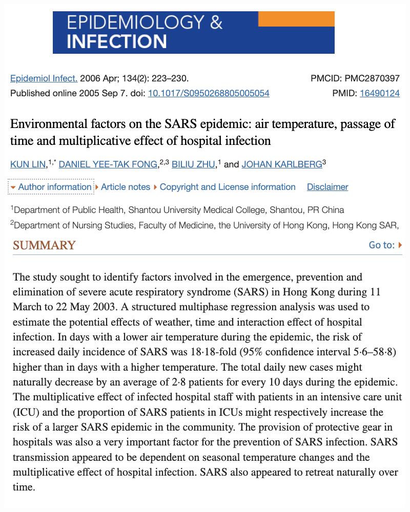
Colder days = 18-fold increase in SARS CoronaVirus infection
Days with colder air temperature resulted in greater numbers of SARS CoronaVirus hospital admissions. This is believed to occur because cold air suppresses interferon production in cells lining the nose, throat and respiratory tract. A interferon is essential to reduce viral growth, any reduction in interferon could potentially increase viral infection.Researchers studied weather conditions in Hong Kong during the SARS CoronaVirus pandemic from March 11 to May 22, 2003. A detailed analysis of records showed an 18.8 fold increase in SARS CoronaVirus patients entering the hospital on days with colder air temperature compared to days of warmer temperatures.
For every 1 degree centigrade increase in air temperature (approximately 2 degrees fahrenheit), there was an "average reduction of 3.6 diagnosed Corona cases per day." (See page 228, paragraph 2)."
Dept of Public Health
Shantou University Medical College
Shantou, China
NOTE: With the average air temperature increasing by about 1 degree centigrade (approx. 2 degrees Fahrenheit) each week in the northern central hemisphere beginning in March - this would suggest a noticeable decrease in infection rates being observed beginning around this time of the year.
Epidemiology & Infection
Vol.134(2):223-230, 2006
Dept. of Public Health,
Shantou Univ. Medical College
University of Hong Kong
View Journal HERE
view PDF
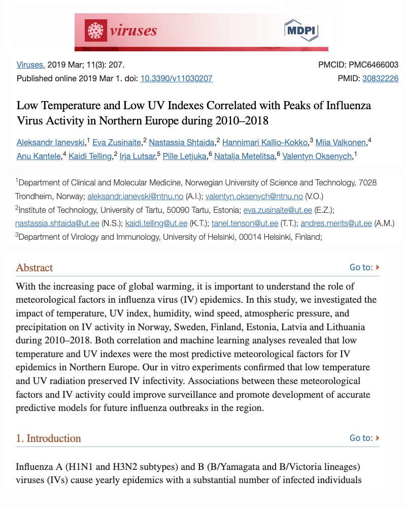
Cold temperature & low sunlight increases flu severity
This study found that influenza in northern Europe was tied to several weeks of colder temperatures and had a more pronounced effect on those 65 and older.Researchers investigated the number of weekly influenza cases reported in six Northern European countries (Norway, Sweden, Finland, Estonia, Latvia and Lithuania) to determine if temperature, sunlight, and humidity could affect the rates of influenza (flu). Researchers concluded that low temperature (especially around 2 degrees C (about 36 degrees Fahrenheit), greatly increased the rate of flu infection. Humidity did not seem to have much of an effect.
In conclusion, researchers stated:
It was shown that low winter temperatures correlated with both influenza incidence and global mortality, which mainly affected those aged 65 and older [21,23,24]. Low UV indexes could also be associated with global mortality. Indeed, data from six Northern European countries reported to the EuroMOMO project showed an excess mortality from all causes between the beginning of Januaries and the end of Februaries in 2010–2018, and coincided with IV (Influenza Virus) epidemic peaks, low temperature and low UV indexes.
Viruses
Vol.11(3):207, March, 2019
Norwegian Univ. of Sci. and Tech.
University of Helsinki
Dept of Immunology
View Journal HERE
view PDF
Common cold virus grows faster in colder temperature
Yale University researchers found the cooler temperatures inside the nasal area (from breathing cold air) reduces the ability of respiratory cells to produce interferon. Why is this important? Interferon is a natural compound that prevents viruses from growing. This provides evidence supporting increased virus growth from colder temperatures.Although not in the coronavirus category, we may be able to learn something from the common cold virus called a rhinovirus. While the temperature of our lungs is typically a warm 98.6 degrees F, (37 C), the temperature inside our nasal cavity is often 7 degrees cooler coming in at around 91.6 degrees F (33 C). This occurs because the nasal cavity is the first tissue exposed to the colder outside air.
As mentioned in the previous section, type-1 interferon is a main defender that protects the human body from the first stages of virus replication. In this 2015 study conducted at Yale University, researchers tested the ability of different temperatures to alter virus growth in mouse airway cells grown in culture (petri-dishes). Their findings showed that warmer temperatures resulted in higher levels of type-1 interferon in nasal cells, which in turn, resulted in far less viral growth. In conclusion, the scientists stated,
These findings demonstrate that in mouse airway cells, rhinovirus replicates preferentially at the lower nasal cavity temperature due, in part, to a less efficient antiviral mechanism. Additional analysis revealed that both type 1 and type 3 interferon were induced at much higher levels at 37 degrees C (98.6 F) than at 33 degrees C (91.6 F) during the rhinovirus replication cycle.
This is another of many published studies showing how warmer temperatures dramatically improve the ability of the human body to fight viruses, and supports findings of lower CoronaVirus infections and severity in warmer temperatures as well.
Yale University School of Medicine
Proceedings Natl Acad Science
Vol.112(3):827-832, Jan 5, 2015
Yale University
Dept. of Immunology
View Journal HERE
view PDF
Body temp of 102 degreees (39C) improves interferon & immune system
Human volunteers placed in hot water baths to raise their core temperature to 102F (39C) showed greatly improved numbers of important immune cells involved in virus removal. Believed to occur from increases in interferon and other important cytokines.Abstract
Induction of hyperthermia (39 degrees C) in human volunteers by immersion in warm water (41-45 degrees C) rapidly alters the cell populations in the peripheral blood. In addition to granulocytosis, there is an alteration of the normal ratios among T-lymphocyte subsets. Following in vitro mitogen stimulation, lymphocytes from hyperthermic individuals produce as much as 10-fold more interferon-gamma (IFN-gamma) than cells withdrawn at basal core temperatures from the same individuals.
A temperature threshold of 39 degrees C for this response suggests potential relevance to fever. No change was noted in the activity of the macrophage population. The possible involvement of interleukin-2 (IL-2) in this enhanced production is discussed. No changes were noted in the circulating levels of IFN-gamma.
Jrnl of Interferon Research
Vol.8(2):143-50
Indiana University
Dept of Biology
View Journal HERE
view PDF
Disinfectants are used with the assumption that they will decrease infection. However, recent studies are showing disinfectants are having an opposite effect and actually increasing rates of influenza and other infections. For example, disinfectant chemicals form inadvertant toxic by-products such as chloroform that weaken the immune system substantially. We'll explain the latest studies documenting these effects.
Common bleach increases (not decreases) child infection rates - toxic disinfectant by-products suspected
Suprising results in this large European study found homes using common household bleach had higher (not lower) rates of child infections. These findings are supported by additional studies demonstrating chlorine bleach reacts with organic substances in water and on surfaces to form immune-suppressing chloroform and others.Common household bleach is typically made from a mixture of water and the chemical sodium hypochlorite. The product is promoted as a "safe" way to disinfectant homes, schools, hospitals and nursing homes. While it is accurate that bleach can reduce surface levels of bacteria and viruses after treatment, scientists are now finding the main chemical in bleach undergoes reactions that form far more toxic chemicals that have not been previously addressed. These chemicals include carbon-tetrachloride and chloroform.
In this study out of Barcelona, Spain, researchers studied homes of over 2000 children ages 6-12 in Spain and Finland. Detailed questionnaires were given to the families inquiring about the use of bleach and frequency of infections including influenza, sinusitis, tonsilitis, ear infections, bronchitis and pneumonia. Results showed that using bleach had an opposite effect of what would be expected and actually increased the rate of influenza by 20% - recurring tonsilitis by 35% - and risk of any infection by 18%. The authors concluded by stating:
"Passive exposure to cleaning bleach in the home may have adverse effects on school-age children's health by increasing the risk of respiratory and other infections. The high frequency of use of disinfecting irritant cleaning products may be of public health concern, also when exposure occurs during childhood."
Centre for Environment and Health
Department of Public Health and Primary Care, KU Leuven, Leuven
Belgium Centre for Research in Environmental Epidemiology (CREAL)
Barcelona, Spain CIBER EpidemiologÃa y Salud Pública (CIBERESP)
Barcelona, Spain University Pompeu Fabra (UPF), Barcelona, Spain
Occup. & Environ Medicine
Vol.73(3), 2016
Department of Public Health
Leuven, Belgium
View Journal HERE
view PDF
Chlorine-based cleaning products create high levels of toxic chloroform
15 common cleaning products containing bleach or other chlorine compounds were tested and found to emit high levels chloroform gas. This is a highly immuno-toxic chemical and adds support to the previous study finding higher rates of respiratory infections among children living in homes using bleach.Researchers tested 15 cleaning products (including household bleach) for levels of toxic chemicals. Chloroform was detected in all 15 products tested and carbon-tetrachloride in 13 products. Both chemicals have been banned and considered highly toxic. After using the products, the estimated exposure for breathing chloroform gas was .041 mg/kg per day and .0240 for carbon-tetrachloride. The scientists concluded by stating:
"According to the calculated result, the total estimated human exposure of chloroform were determined to exceed the tolerable concentration of inhalation exposure presented by the World Health Organization."
This study adds further support to the hypothesis that chemical disinfectants, rather than helping, could be a significant driving factor in worsening the current state of COVID-19 infections via a weakening of the human immune system. Because of the widespread increase in residential chemical disinfectant use since the COVID-19 outbreak, it is imperative to investigate the immuno-toxicology of chemical disinfectants with special emphasis on children, the elderly and those with weakened chemical detoxification systems (i.e. cytochrome P-450). Non-toxic disinfectants being shown to have similar effectiveness to chemical disinfectants should also be encouraged - including basic detergent, low ph white vinegar and thymol.
Chemosphere
Vol.174:157-164, May, 2017
Dept of Environ. Education
Kongju National University
Republic of Korea
View Journal HERE
view PDF
Toxic gases created after using household bleach - Health Concerns
8 chlorine bleach products were tested and found that when contact was made with surfactants (such as soap or detergent), that it created high levels of the chemicals chloroform and carbon tetrachloride. These chemicals are highly toxic to the immune system and liver, thereby potentially increasing infections.Sodium hypochlorite is the main disinfectant chemical found in household bleach. This study demonstrated that a number of new and highly toxic chemicals are found in indoor air immediately following the use of bleach to disinfect.
Researchers tested 8 different chlorine bleach products for their ability to emit or create potentially toxic chemicals. Indoor air samples were taken before, during and 30 minutes after bathroom, kitchen, and floor cleaning applications. Of great concern, the highly toxic chemicals chloroform and carbon-tetrachloride significantly increased during the use of bleach containing products.
Researchers went on to state:
"One of the most surprising results was the presence of carbon tetrachloride (a probably human carcinogen and powerful greenhouse gas that was banned for household use by the U.S. Food and Drug Administration in very high concentrations (up to 101 mg/m(-3). By mixing surfactants or soap with NaOCl (bleach), it was shown that the formation of carbon tetrachloride and several other halogenated VOCs (15 in total), is possible.
Environmental Science & Tech.
Vol.42(5):1445-51, 2008
Dokuz Eylul University
Buca, Ismir, Turkey
View Journal HERE
view PDF
Interferon decreased by chemicals in bleach
Interferon in our lungs, throat and nasal area is essential in stopping viral infections literally before it starts. Unfortunately, common household bleach disrupts the ability of interferon to function properly, which provides an explantion why other studies have found higher infection rates among children living in homes using bleach.Since it is now well established that interferon production by cells lining the inside of our nose, throat and lungs is essential for preventing viral infections (including COVID-19), the next question is if there are environmental circumstances that can reduce interferon output?
In this next study from the Dept. of Microbiology and Immunology at University of Louisiana School of Medicine, researchers pretreated mouse embryo cell cultures with a number of different chemicals. Results showed pretreatment with the chemical chloroform (which is created while using bleach) severely inhibited the production of alpha/beta interferon by cells.
Since chloroform gas was shown to reach exceptionally high air levels after use of bleach products (as demonstrated in a previous journal review), this study provides a plausible explanation for the higher rates of influenza (and other infections) in 6-12 year old children living in homes using bleach products.
Environmental Research
Vol.3192:355-361, Aug, 1983
Dept of Microbiology & Immunology
University of Louisville, Kentucky
View Journal HERE
view PDF
Bleach damages DNA in immune cells
Lymphocytes are the #1 immune cell protecting us from COVID-19. However, common bleach caused significant damage to the DNA of lymphocytes. Since DNA is what controls the cell's function, this would suggest bleach has the ability of reduce the function of these important cells.Our lymphocyte white blood cells rely on their DNA to tell them what to do and how to stop a virus. In this study, it was found that common bleach damaged the DNA in these white blood cells.
Lymphocytes are the type of white blood cell found in abnormally low numbers in people who develop severe CoronaVirus and have been found low in those who also do not survive Influenza. In this study, researchers wanted to determine how different levels of the chemical sodium hypochorite (the main chemical in common bleach and some water treatments) affected the chromosomes of lymphocytes. Blood samples were collected from healthy non-smoking adults and added to a culture that encourages the survival and growth of lymphocytes. Blood cultures were exposed to different concentrations of sodium hypochlorite at levels measured in micrograms per milliliter. Levels included - 0.030 - 0.065 - 0.100 - 0.25 - 0.50 - 1 - 2 and 4.
In summary, the researchers stated (page 117):
A significant increase in chromosomal aberration (abnormality) frequency was observed in all treatments of NaOCl (sodium hypochlorite).... at 24 and 48 hours compared with the negative control...
In conclusion, our data provide evidence that NaOCl (sodium hypochlorite) increases the chromosomal aberration rate, micronuclei formation (small nucleus) and cytotoxicity (cell damage) in human lymphocyte cells in vitro at concentrations approximate 33 times lower than that found in drinking water.
Department of Biology, University of Kafkas, Kars, Turkey
Cytotechnology
Vol.59(2):113-119, March, 2009
Department of Biology
University of Kafkas, Turkey
View Journal HERE
view PDF
Birth defects higher from common disinfectant
The second most common disinfectant after bleach is called Quaternary Ammonium Compounds. These have also been found to cause severe health consequences including neural tube birth defects. The "neural tube" is the very first stage of brain growth in humans and other mammals.Another group of disinfectants is called Quaternary Ammonium Compounds. These chemicals are used in common commercial and residential disinfectants and do not contain bleach. Researchers at the Edward Via College of Osteopathic Medicine in Virginia became interested in these chemicals after being informed of two separate incidents at two laboratories in which there was an observed decrease in animal health and an increase in neural tube defects after switching to a particular disinfectant. The disinfectants in question contained a combination of two chemicals including alkyldimethylbenzyl ammonium chloride (ADBAC) and didecyldimethyl ammonium chloride (DDAC). To determine if the disinfectants were causing a problem, both mice and rats were divided into groups with some being fed the chemicals, other receiving injections and others being exposed to the chemicals normally in the ambient air. The main findings showed exposure from the ambient air (breathing in fumes) caused more neural tube defects than the other exposure conditions. Also of concern, the rate of neural tube birth defects continued for 2 generations, (thereby suggesting potential for genetic damage). In conclusion the researchers stated,
These results demonstrate that ADBAC+DDAC in combination are teratogenic to rodents. Given the increased use of these disinfectants, further evaluation of their safety in humans and their contribution to health and disease is essential.
Although this study is not related specifically to the CoronaVirus (or other viral infections), it is of concern as this type of disinfectant is being used with much greater frequency during the COVID-19 situation.
Birth Defect Research
Vol.109(14)1166, 2017
E. VA College Osteopathic Med
VA-MD College Veterinary Med
Washington State University
View Journal HERE
view PDF
Detergent as effective as disinfectants for avoiding infections
Studies comparing chemical disinfectants to detergents found using basic detergents was just as effective for reducing infections in hospital patients.As seen in the previous reports, common chemical disinfectants are suspected of weakening the the immune system, thereby resulting in higher infection rates for influenza and other pathogens. In this report out of Freiburg University Hospital in Germany, researchers reviewed 4 scientific studies investigating the effectiveness of chemical disinfectants for reducing infection in a hospital setting.
After analyzing results, the study concluded that cleaning of floors with basic detergents was just as effective as using chemical disinfectants.
Scientists concluded by stating:
"None of these studies showed lower infection rates associated with routine disinfection of surfaces (mainly floors) versus cleaning with detergent only... Disinfectants may pose a danger to staff, patients, and the environment and require special safety precautions. However, targeted disinfection of certain environmental surfaces is in certain instances an established component of hospital infection control. Given the complex, multifactorial nature of nosocomial (hospital) infections, well-designed studies that systematically investigate the role of surface disinfection are required."
American J Infectious Control
Vol.32(2):84-89, 2004
Inst. of Env. Med. & Hosp Epidem.
Freiburg University Hospital, Germany
View Journal HERE
view PDF
Trends identifying annual declines in immune system white blood counts in the U.S. and other populations are of particular concern. Any continued decline (as currently being noted annually) clearly predicts dramatically increased viral infections in the near future. Only by reversing this trend will viral infections decline. It is hoped this study will bring needed attention to this important topic.
Immune system counts dropping annually in the U.S.
Major study out of the National Institutes of Health showing white blood counts have been dropping annually in adults over the past 50 years. Since having low numbers of white blood cells is well known to increase the frequency and severity of viral infections, this trend serves as a wake-up call and is reason for serious concern. The importance and meaning of these numbers are discussed along with references to environmental factors found to decrease white blood cell counts.Not Good News... The part of our immune system that plays the biggest role in removing viruses from the body has been steadily declining for more than four decades. These virus fighting cells (called white blood cells), are dropping at the rate of about 50 per year in each cubic millimeter of our blood (a little less than a drop). These were the findings reported from the National Institutes of Health (NIH) Aging Division, which has been looking at blood test reports over the past 40 years.
Counts Below 3,500 = Higher Risk of Death
When you go to the doctor for a routine blood test, they will often take a blood sample and measure your white blood count. This is the number of immune system white blood cells in a cubic millimeter of blood (a little less than a drop). The so-called "normal range" is what 95% of the population scores, with 2.5% above that range and 2.5% below that range.
Normal Range Shifting
The NIH study looked at 2,853 people spanning from 1958 to 2002. Participants were asked to return every few years to conduct cognitive and blood tests, thereby observing changes in the aging process. Along with the steady decline in blood counts seen in participants each decade, scientists found people with white blood counts below 3,500 (the number considered very low) had a 3-times greater risk of dying than people who averaged closer to 5,000 and 6,000. This certainly makes sense, since having lower numbers of immune-system fighting cells should correspond with a faster growth rate for viruses.
Extremely Low Blood Counts Increasing Rapidly
Each column in the chart above gives the number of people per 1000 with a white blood count below 3,500 (the number considered low). In the first decade from 1958-1969, people with very low white blood counts averaged on 2 per 1000 people. During the second decade (1970-79), the number of people with very low white blood counts doubled to 4 per thousand. In the third decade (1980-89), this number nearly doubled again reaching 7 per thousand people. Of great concern, during the fourth decade (1990 to 2000), there was a tripling of the number of people with low white blood counts - now reaching 22 people per thousand!
The study was published in 2007, and does not have numbers for the last two decades from 2000 to 2020. Because of the exponential growth (and extreme seriousness of this condition), it would be in our interest to determine how numbers have changed during this last 20 year period - especially in light of new research showing that having a low white blood count greatly increases the risk of overall death including a fatal outcome in influenza and apparently, in CoronaViruses as well.
The average white blood count over the 42 year study was also seen to be dropping for those involved in the study. While scientists stated that reductions in cigarette smoking was partially responsible (cigarette smokers have higher counts), they also stated that smoking did not explain all of the reductions. Below is the chart that shows the reductions in average white blood cell count from 1958 to 2002.
NIH graph
Why are Blood Counts Dropping?
Obviously, such dramatic changes in blood counts over a relatively short period of time suggests something around us is suppressing white blood cell development (which occurs in our bone marrow, lymph organs and thymus). Toxicology research over the past several decades has identified a number of circumstances in daily life that can lower the number of white blood cells and/or quality.
Studies investigating environmental factors that lower white blood counts will be discussed at the end of this report and include chemicals that can suppress bone marrow production of cells, chemicals found to damage DNA inside white blood cells, as well as the myriad of chemicals found to weaken and damage the thymus gland. The thymus is critically important for the final maturation of white blood cells called "T-Cell lymphocytes." These cells play a critical role in organizing and continuing the attack against viruses in the body. Perhaps not coincidentally, individuals who eperience severe reactions to COVID-19 have extremely low levels of T-Cells. Also of interest, the thymus plays an essential role in filtering/removing autoimmune cells causing arthritis, lupus, and other autoimmune diseases.
J of Am Coll. Cardiology
Vol.49(18):1841, 2007
National Institutes of Health
Baltimore, MD
Lead Author: Dr. C. Ruggiero
View Journal HERE
view PDF
Toxicology is the branch of medical science that investigates how circumstances around us can damage human health. Going far beyond cigarettes or alcohol, here we summarize studies that have identified specific chemicals in consumer products (previously considered safe) and how they are able to cause serious weakening of specific immune system cells needed to protect from COVID-19. In fact, hundreds of research centers around the world (including Harvard University) are now stating "immuno-toxicology" appears to be the primary reason for far higher rates of COVID-19 infection in industrialized countries. These will be discussed below but excludes "air polllution" which has its own category.
Common flame retardant causes major weakening of natural killer cells
A very common flame retardant (called TBBPA) was shown to nearly shut down the ability of natural killer (NK) cells to perform their job in killing cancer infected cells. Because NK cells function in similar fashion to killing viruses, this study is important to understand.TBBPA is a common flame retardant used in 90% of circuit boards in computers and other electronic products. It is also added to reduce the burning speed of plastic, clothing, carpets, upholstery and even found in polycarbonate drinking bottles (which then leaches into the water). It evaporates into the air and accumulates onto dust which is then inhaled by occupants of the home. One study showed dust levels of flame retardants in U.S. homes was 10 times higher than in homes in the European Union. Policy differences on allowable levels of flame retardants is believed to explain these differences.
Levels of the flame retardant TBBPA in human blood range from 0.24-0.71 ng/g in Norway to an average of 4.5 ng/g in some Japanese populations.
In this study from the University of Tennessee, researchers were interested whether this flame retardant could affect human immune system function. They took blood from healthy volunteer donors and exposed their natural killer (NK) cells to different levels of TBBPA for either 24 hr, 48 hr, or 6 days. NK cells were then tested for their tumor killing ability, their ability to bind (attach) to cancer cells and ATP levels (which is a measurement of energy produced in the cell by mitochondria).
KILLING ABILITY WAY DOWN
NK cells lost their ability to kill cancer cells at even the lowest levels tested. For example, NK cells exposed to TBBPA for 24 hours at levels of 5 μM caused a 96% reduction in the number of cancer cells killed by NK cells. A 24 hour exposure to 1 μM of TBBPA caused a 39% reduction in NK tumor killing ability. Even at the lowest 0.5 μM level, NK cells lost 23% of their killing ability.
BINDING ABILITY WAY DOWN - 24 Hour Exposure
For an NK cell to kill a cancer or virus infected cell, it must first attach onto the cell using special receptors. TBBPA reduced their binding ability as well. While no binding loss was seen from the 1 μM dose, there was a 19% loss of binding ability at 2.5 μM and 70% loss of binding at 5 μM in the 24 hour experiment.
6 DAY EXPOSURE
NK cells were also exposed to very low levels of TBBPA in the 6 day experiment, going down to as low as 0.05 μM. All exposure levels except for the 0.05 level caused a significant reduction in their tumor-cell-lysing (killing) function.
TBBPA caused the following decreases in NK cancer cell killing function:
0.1 μM TBBPA caused a 20% loss of NK cancer killing ability
0.25 μM caused a 33% loss of cancer killing ability
0.5 μM caused a 54% loss
1 μM caused an 89% loss of killing ability
This low level exposure also caused a significant decrease in binding ability.
Below are direct comments from the study authors:
[BEGIN QUOTE]
1. Chemically-induced diminution of human NK cell immune function could leave an individual vulnerable to malignant neoplasias and/or viral infection.
2. Previous studies have indicated that humans with a defect that leaves their NK cells unable to lyse target cells have a predisposition to development of certain tumors (Ortaldo et al., 1992)
3. Further, the ability of an initial exposure to TBBPA to cause lasting and/or ensuing
disruption of lytic (killing) function was examined, as a lasting disruption of NK function by a brief encounter with TBBPA would indicate a greater level of potential harm from exposure to this compound.
4. A 24 hr exposure to as little as 0.5 μM TBBPA caused a significant loss of lytic function in NK cells. When the concentration was 5 μM there was a nearly complete loss of lytic function at 24 hr.
5. Previous studies have shown that a variety of environmental contaminants cause decreases in NK cell function that persist once the compound is removed. These include another brominated flame retardant, hexabromocyclododecane (HBCD) (Hinkson and Whalen, 2009), several organotin compounds (tributyltin [TBT], dibutyltin [DBT], and triphenyltin [TPT]) (Whalen et al., 2002; Dudimah et al., 2007a,b; Whalen et al., 2003) an organochlorine compound, pentachlorophenol (PCP) and a carbamate compound, ziram (Taylor et al., 2005).
[END QUOTE]
This last statement of many contaminants having the ability to reduce NK function is concerning as it suggests there may be a synergetic effect of chemicals coming together to cause more harm than from single exposures.
Journal of Immunotoxicology
Vol.6(4):285-292, Dec 2009
Department of Chemistry
Tennessee State University
View Journal HERE
view PDF
Monosodium Glutamate (MSG) damages immune cells
Our spleen is a major immune system organ storing lymphocytes that remove bacteria and viruses. Researchers state the food additive MSG generated nearly a 30% increase in pro-inflammatory cytokines and over 35% inrease in Reactive Oxygen Species (ROS) in immune cell lymphocytes stored in the spleen.ABSTRACT
Globally, the trend of using food additives and eating ready-made fast food has led to a deleterious impact on immune organs. Monosodium glutamate (MSG), as a food additive in a high-lipid diet (HLD), acts as a silent killer of immune cells. Hence, the present study aimed to evaluate the role of MSG in HLD on spleen injury in rats. Results showed that a 2.52-fold and 1.91-fold increase in spleen index in MSG and MSG + HLD group indicates splenomegaly, whereas a 1.36-fold and 1.29-fold increase in pro-inflammatory cytokines in MSG and MSG + HLD-fed rats, respectively, promote the inflammatory response. Additionally, MSG and MSG + HLD induce oxidative stress by 1.81-fold and 1.1-fold increased generation of reactive oxygen species (ROS) in macrophage population, and 1.38-fold and 1.36-fold increased generation of ROS in lymphocytes population, respectively. Furthermore, mitochondrial membrane potential was significantly reduced by 1.43-fold and 1.18-fold in MSG and MSG + HLD groups. Therefore, the current study argues that MSG has more detrimental effects on the spleen than MSG + HLD due to the presence of antioxidants in HLD, which suppresses the deleterious impact of MSG. Hence, it can be inferred that MSG induces spleen injury via targeting redox-guided cellular signaling with inflammatory response, leading to severe immune system anomalies.
Open Life Sciences
Vol.17(1):22-31, Jan 29 2022
Department of Physiology
Serampore College, India
View Journal HERE
view PDF
Harvard identifies 34 chemicals suspected of increasing COVID-19
34 chemicals were identified that appear to mimic and disrupt important immune system communication signals known as cytokines. Any disruption of cytokine communication between immune cells could potentially turn an asymptomatic or mild infection into a severe or fatal infection.Below are five classes of endocrine disrupting chemicals Harvard researchers identified which they believe could be weaken immune function and worsen viral infections.
CHEMICAL #1 ON THE LIST - PCB'S
Found in the coloring of common housepaint and sometimes very high in homes built in the 60s and 70s, because of its use in caulking, paint and wood varnish. It's also emitted from coal-fired power plants which then rain down in our waterways, soil and pastures where it concentrates in fish and even reaches high levels in meat of pasture raised cattle.
CHEMICAL #2 - DIOXIN
Found in bleached paper, car exhaust, and emitted at high levels in backyard trash burning and municipal incinerators. Also forms in some pesticides and emitted from coal-fired power plants.
CHEMICAL #3 - PESTICIDES
Cytokine immune disruption potential was also found from a number of common pesticides used in food and homes. This included the pesticides chlordane (found in homes before 1990), chlorpyrifos (in food and newer homes), atrazine (weed killer), cypermethrin, DDT and DDE (which we ingest from foods grown imported from South America that allow DDT).
Since several of these pesticides are found at high levels in foods produced using chemical-based agriculture, it would suggest eating organic could have a highly protective effect from Covid-19. Some studies have shown high Covid outbreaks near agricutural communities which would seem plausible as well.
CHEMICAL #4 - PLASTICS
When looking through Harvard's cytokine damaging list - I also see mention of our most common plastic chemical used in water bottles, food packaging and even dental fillings. This includes bisphenol A (BPA), bisphenol F (BPF), and the new BPA replacment bisphenol-S (BPS). This is telling us that even BPA-Free is meaningless as replacement plastics are typically BPS. So, the next time you drink from a water bottle, you might want to question why you are choosing to drink chemicals Harvard scientists say allow Covid-19 to grow faster in your body.... (great huh?)
CHEMICAL #5 - DISINFECTANTS
Certainly an irony of ironies. It now appears that the disinfectant and antibacterial chemicals we have previously believed would prevent infection are now showing they can increase the risk of infection.
One of these disinfectant chemicals found to fool the immune system communication cytokines is "triclosan," and while banned in some products, it's still used in antbacterial lotions, liquid hand soaps, foaming hand anitizers, mouthwash, deodorants, paints and even wooden pencils (which I personally loved to chew as a middle school student - you ever do that?).
CHEMICAL #6 - COSMETICS
Cosmetics such as facial makeup and lipstick need to be thickened - and the syrupy concoction known as phthalates does the trick. Unfortunately, that's also on Harvards NO-NO list of immune system chaos. Cosmetics also need preservatives and contain chemicals called parabens. Yep, that's on their list too.
Environmental International
Vol.152:106232, Dec 2021
University of Paris
School of Public Health
Harvard Medical School
View Journal HERE
view PDF
Harvard scientists link severe COVID-19 with common chemical in home products
The chemical of concern is PFAS and has been used in consumer products for over 50 years. When comparing patients with mild, moderate and severe COVID-19, as the severity increased, so did the likelihood of testing positive for PFAS. The chemical has recently been found highly toxic to the immune system.INTRODUCTION
PFAS chemicals are added to consumer products to make them resistant to water and oil. They are added to pizza boxes, microwave popcorn bags, frozen microwave meals, paint and are very common in cosmetics, lipstick and sunscreens.
Recent studies have shown PFAS chemicals decrease numbers of natural killer cells (our main Covid defense) as well as decrease antibody production.
BELOW IS A SUMMARY OF THIS STUDY PUBLISHED IN THE HARVARD SCHOOL OF PUBLIC HEALTH NEWSLETTER
People who had elevated blood levels of a toxic chemical called perfluorobutanoic acid (PFBA) had an increased risk of a more severe course of COVID-19 than those who did not have elevated levels, according to a new study led by Harvard T.H. Chan School of Public Health.
PFBA is part of a class of man-made chemicals known as perfluorinated alkylate substances (PFASs), which have previously been shown to suppress immune function.
The study, published December 31, 2020 in PLOS ONE, was led by Philippe Grandjean, adjunct professor of environmental health.
PFASs have water- and grease-resistant properties and are used in a wide variety of products, including nonstick cookware, waterproof clothing, food packaging, and firefighting foams. PFBA, more than other PFASs, is known to accumulate in the lungs, according to the study.
Researchers looked at PFAS levels in blood samples from 323 Danish individuals infected with the coronavirus. They found that those with higher PFBA levels had higher odds of being hospitalized, winding up in intensive care, and dying than those with lower levels.
The findings suggest that further study is needed to determine whether elevated exposures to other environmental immunotoxicants may worsen COVID-19 outcomes, the authors wrote.
PLOS ONE
Vol.15(12):e0244815
Harvard School of Public Health
Univ. Sothern Denmark
View Journal HERE
view PDF
Chemicals in cosmetics & plastics weaken immune system
Another disinfectant chemical often used in cosmetics is being found to damage tissues involved in development of white blood cells. The chemical phenol damages the lymph systems (tonsils, lymphnodes, etc) that make lymphocytes.The chemical phenol is used in many products around us. In this study from the Dept. of Basic Sciences, University of Tehran, researchers found that phenol caused severe deterioration of organs that make lymphocyte white blood cells (lymphocytes are typically extremely low in fatal COVID-19 situations).
Phenol is used to make plastic bottles (bisphenol-A), epoxies, nylon, detergents, and a number of weed killers. It is used at a 1.4% concentration in sprays for sore throat (Chloraseptic) and is also used as a preservative in some vaccines. [Wikipedia].
Phenol derivatives are also used in the preparation of cosmetics including sunscreens, hair colorings, skin lightening preparations, as well as in skin toners/exfoliators. Because of its anesthetic and disinfectant properties, phenol is used widely in pharmaceutical products such as ointments, ear and nose drops, sprays and antiseptic lotions (Finkelstein et al. 2007).
Once phenol enters the blood it is transferred to the bone marrow (where all immune cells originate) where it is then converted into several other biologically toxic compounds. Due to safety concerns, phenol is banned from use in cosmetic products in the European Union and Canada - but is still allowed in cosmetics in the U.S. (Wikipedia).
How Phenol Weakens the Immune System
Our lymphocyte white blood cells are the main fighters that remove viruses from the body. They begin in the bone marrow and then develop further in other lymphoid organs including thymus, spleen, lymph-nodes, tonsils and bone-marrow. In this study, conducted at the Department of Basic Sciences, University of Tehran, researchers found that phenol administered to mice at levels of 80, 180 and 320 mg/kg/day, resulted in a visual decrease in the quality of the spleens, thymus, and other lymphoid organs. This included a reduced number of thymocytes in the thymus (thymocytes are cells that eventually become mature lymphocyte T-Cells - and were found deficient in those experiencing severe COVID-19 symptoms). In a study by Baj et al. (1994), it was reported that subjects with the highest levels of phenol in urine had decreased T-helper lymphocyte numbers. Since these organs are essential for lymphocyte immune cell development, this would strongly imply that phenol is another chemical to be added to the growing list of chemicals with the ability to weaken a critical part of our immune system.
Comparative Clinical Pathology
Vol. 23(3): 529-534, 2014
Dept. of Basic Sciences
University of Ilam, Iran
View Journal HERE
view PDF
Electromagnetic exposure weakens immune system
Electromagnetic fields are produced by electric devices around us. In this study - 15 men and women working near a large power cable in their office were found to have lower levels of immune cells called natural killer cells (these are critically important in fighting viruses).The objective of this study was to analyze the immune response to electromagnetic fields (ELMFs) in seven men and eight women employed in a museum. All individuals worked in a room that contained a building power cable running along the back wall behind their work stations. This created a 50 Hz exposure in the range 0.2-3.6 microT and 40-120 Volts per meter. These are levels considered acceptable for people in the workplace. Their blood test results were compared to a similar age control group of 45 subjects.
Results showed the electromagnetic exposure had significant effects on reducing immune system function. Women were found to experience a significant reduction in the percentage of B-Cell lymphocytes (cells that make antibodies) and also natural killer cell lymphocytes. Women also showed significantly lower levels of interferon gamma (which helps stop viruses before they start).
Men exposed to the electromagnetic fields experienced a statistically significant reduction in both the number and percentage of natural killer cell lymphocyte subsets.
In conclusion, the researchers stated,
"On the whole, this investigation demonstrates a reduction of blood NK (Natural Killer) lymphocytes and of the production of interferon gamma in workers exposed to low frequency ELMFs."
Science of Total Environment
Vol.270:13-20, 2001
Dept of Medicine and Sciences
Univ. of 'G.D'Annunzio, Italy
View Journal HERE
view PDF
Extremely low frequency electric fields damage important Covid immune defense
While living close to cell towers has been shown to decrease immune function, another type of electromagnetic radiation causing concern is called Extremely Low Frequency Radiation (ELFR). This is emitted by overhead power lines and typical home AC electric wiring.Abstract
Extremely Low Frequency-Magnetic Fields (ELF-MF) are possible carcinogens to humans and some data suggest that they can act as promoters or progressors. Since NK cells play a major role in the control of cancer development, an adverse effect on ELF-MF on NK function has been hypothesized. We examined NK activity in 52 workers exposed to different levels of ELF-MF in various activities. Individual exposure was monitored during 3 complete work-shifts using personal dosimeters. Environmental exposure was also monitored. ELF-MF levels in the workers were expressed as Time-Weighted Average (TWA) values. NK activity was measured in peripheral blood lymphocytes (PBL). In the whole group the median occupational TWA was 0.21 µT. According to the TWA levels, workers were classified as low exposed (26 subjects, TWA ≤ 0.2 µT) and higher exposed workers (26 subjects; TWA > 0.2 µT). In higher exposed workers, we observed a trend to reduce NK activity compared to low exposed, but the difference was not significant. Then we selected a subgroup of highest exposed workers (12 subjects; TWA > 1 µT); no difference was observed between low and highest exposed subjects in the main personal variables. Considering both E:T ratios from 12:1 to 50:1 and Lytic Units, a significant reduction in NK activity was observed in the highest exposed workers compared to the low exposed. Multivariate analysis showed a significant negative correlation between exposure and LU, while no correlation was evidenced with other personal characteristics. ELF-MF are considered possible carcinogens, and existing data suggest that they can act as promoters. Due to the role of NK activity in host defence against cancer, the results obtained in this study in workers exposed to ELF-MF levels exceeding 1 µT are in agreement with this hypothesis, and support the need for further investigation in this field.
Science Total Environment
Vol.407(3):1218, Jan 2009
Univ. of Medena & Reggio Emilia
Italy
View Journal HERE
view PDF
Living near cell towers reduces immune cells
People living near cell towers were found to have reductions in important immune system cells and lower levels of antioxidants when compared to people living farther away.Microwave radiation emitted by mobile phone base stations have raised concerns on its adverse impact on humans residing in the vicinity of mobile phone base stations. This present study (conducted by the Cancer and Radiation Biology Laboratory, Mizoram University, India) compared blood test results from 40 people living less than 30 meters from a cell tower with 40 people living more than 300 meters away (nearly 1000 ft). Both groups were matched according to age, gender, dietary pattern, smoking habit, alcohol consumption, duration of mobile phone use and average daily mobile phone use.
The first abnormality detected increased frequency of what is called "micronuclei" in people living close to the cell tower. Micronuclei were found in lymphocyte white blood cells and are evidence of DNA damage. This is of concern since lymphocytes are the primary blood cell that removes viruses from the body. Any reduction in lymphocyte numbers and quality would be expected to increase the onset and severity of any viral infection.
The second abnormality detected in people living close to a cell tower was a "significant reduction" in blood level antioxidants. This included reductions in glutathione, superoxide dismutase, and catalase. These 3 antioxidants provide important protection to all cells by continuously scavaging free-radicals that could otherwise damage the cell well, cell DNA and cellular mitochondria - eventually resulted in cell death. Free radicals are formed from basic oxygen metabolism as well as exposure petroleum-based chemicals. They are also formed in high levels as a consequence of the immune-system ridding the body of a virus infection. Therefore, having low levels of antioxidants would be expected to increase collateral damage to healthy cells during any viral infection.
Electromagnetic Biol. Medicine
Vol.36(3):295-305, 2017
Dept of Zoology - Cancer & Radiation
Mizoram University, India
View Journal HERE
view PDF
Car exhaust and electromagnetic fields combine to weaken immune system
This study found simultaneous exposure to both vehicle exhaust and electromagnetic fields (ELMFs) resulted in lower number of immune system natural killer cells.In the "real-world," people are not exposed to harmful situations one at a time. In this study, women with and without allergies were exposed to both ELMFs and vehicle exhaust to determine how single and combination exposures affected their immune system. While most lymphocytes were not affected with the levels studied, it was found that exposure to ELMFs reduced interferon (a natural chemical that enhances immune activity) and decreased natural killer cell function.
In conclusion, the researchers stated,
"This result, which demonstrates a different influence of ELMF on non-atopic (non-allergic) fertile women exposed to the toxic compounds of traffic and the non-atopic (non-allergic) ones, suggests that exposure to toxic compounds may increase the effects of ELMF in the atopic (allergic) subjects more than in those non-atopic. (non-allergic)"
Industrial Health
Vol.38(3):294-300, July, 2000
Dept of Science
University G.D Annunzio
Italy
View Journal HERE
view PDF
Common pesticide weakens primary defense against CoronaVirus
A common food pesticide was found to weaken an immune system cell that plays a critical role in protecting from a major CoronaVirus infection. The cells are called natural killer cells and were found to be highly susceptible to damage from the pesticide known as chlorpyrifos.When a CoronaVirus is on the verge of starting a major infection in the human body, an immune cell called a natural killer cell is attacking the virus with intensity and desperately trying to sway the see-saw so the virus loses and the body wins. Unfortunately, the common food pesticide chlorpyrifos, appears to have the ability to sway things in favor of the virus winning.
In a study out of Nippon Medical School in Tokyo, Japan, researchers exposed natural killer cells to low levels of chlorpyrifos from 0 to 100 parts per million for 1 to 72 hours. The effect was so great that the cells didn't just become "weak" - the exposures resulted in the death of natural killer cells by a process called apoptosis.
Toxicology
Vol.239(1-2):89-95, 2007
Nippon Medical School
Tokyo, Japan
View Journal HERE
view PDF
Fragrance chemical weakens virus defense
A common chemical used in fragrances, cosmetics and plastics was shown to reduce levels of a very important virus fighting part of your immune system called interferon type-1.The chemical in question is known as phthalates (pronounced thal-ates) and is used as a solvent in perfumes/colognes - plug-in air fresheners - as a thickening agent in cosmetics - lotions - sunscreens - and also used to make plastics soft and pliable.
The news is not all good, researchers at the College of Medicine in Taiwan found this common chemical also has the ability to disrupt the secretion of interferon type-1. The cells affected are important cells lining our respiratory tract called dendritic cells. Dendritic cells are one of the first responders in virus attacks and work as a cheerleader, sending out chemical messages to motivate other cells to join the attack against viruses. One of these motivational chemicals is called interferon type-1, which calls in and stimulates a number of important white blood cells (including natural killer cells.
Unfortunately, the ability of dendritic cells to produce interferon is reduced significantly by the presence of the phthalates. Without sufficient levels of interferon type-1, there is an inadequate immune response to the first viruses entering into the respiratory tract.
Researchers concluded by stating that phthalates may interfere with immunity against infection, and can weaken the ability of dendritic cells to stimulate other important immune cells to join in the fight against the virus. The chemical does this, not by causing DNA damage, but instead, by suppressing critical genes that control interferon output.
Allergy
Vol.68(7):870-09, 2013
Kaohsiung Medical University
Taiwan
View Journal HERE
view PDF
Food Coloring Decreases White Blood Count
Titanium dioxide is food coloring used in many food products and medicines to make the item appear white in color. This study showed titanium dioxide caused strong reductions in immune system function including decreased white blood count.Mice were exposed to the titanium dioxide particles at levels of 0, 1.25, 2.5, or 5 mg kg-1 body weight for 9 consecutive months. After the 9 month period, animals were tested for many aspects of blood and immune system quality. Results showed the color accumulated in the thymus gland which is the organ responsible for lymphocyte T-Cell development. Reductions in many subsets of lymphocytes were noted, including antibody producing B-Cells, T-helper Cells and natual killer cells.
Of interest, as mentioned in studies from Section 1, T-helper and natural killer cells were found to be low in patients with CoronaVirus infections. This study gives evidence that common food colorings have the ability to contribute to a weakening immune system.
Environmental Toxicology
Vol.32(10):2234-2243, 2007
Laboratory of Food Safety
Huaiyin Normal University
China
View Journal HERE
view PDF
4 food preservatives damage DNA in white blood cells
The food additives citric acid, phosphoric acid, benzoic acid and calcium propionate were found to cause DNA damage in white blood cell lymphocytes.To prevent spoiling, foods often contain chemical preservatives. In this study, immune system lymphocytes were exposed for 1 hour to four different food preservatives: Citric acid at a level of 200 micrograms/milliliter - Phosphoric Acid at 25-200 micrograms/ml - Benzoic Acid at 50-500 micrograms/milliliter and Calcium Propionate also at 50-100 micrograms/milliliter.
Results showed that the levels tested were able to induce DNA damage.
The most toxic preservative was phosphoric acid (found in popular soft-drinks) while calcium propionate caused the least amount of DNA damage. It is important to note that when DNA damage occurs, the immune system cell will not function properly.
Toxicology & Industrial Health
Vol.30(10):926-37, 2014
Faculty of Health Sciences
Akara University, Turkey
View Journal HERE
view PDF
This next section includes peer-reviewed articles suggesting new treatment options for COVID-19. As those with COVID-19 or Long Covid have clearly identifiable abnormalities in immune function (i.e. low natural killer cells and/or high autoantibodies), it would seem plausible to apply interventions that could reverse these contditions, thereby improving immune and defense systems. These will be discussed below.
Antioxidant pycnogenol prevents Covid-19 damage by removing free-radicals
A common factor identified in moderate and severe COVID-19 patients is reduced levels of protective antioxidants such as glutathione. The pine bark extract pycnogenol functions to "take the place" of low antioxidants such as glutathione, thereby reducing tissue damage and immune suppression.When the SARS coronavirus enters cells, it results in formation of large amounts of free-radicals. This can potentially lead to significant damage to blood vessels and other tissues.
Normally, cells in the human body have a defense against these free radicals, including production of antioxidants (such as glutathione) which neutralize excessive free-radicals.
However, those with moderate to severe COVID-19 have been found to have far lower levels of these antioxidant defenses, thereby allowing free-radicals to cause more harm, and providing an explanation for their more severe reaction to the virus.
To compensate for this lack of antioxidant protection from COVID-19 free-radical formation, scientists are suggesting the administration of the over-the-counter supplement called Pycnogenol, which is made from French Maritime Pine Bark.
The logic here is that pynogenol will function to reduce the excessive formation of free-radicals causing oxidative stress, inflammation and damage to cells in the body.
Int J Antimicrobial Agents
Vol.56(6), Dec 2020
Horphag Research, Switzerland
View Journal HERE
view PDF
Vitamins used in treatment of COVID-19
Those who do become infected with COVID-19 are being found to have low levels of important vitamins - suggesting this as a causal factor. A number of vitamins have been reported to be highly beneficial in preventing and/or reducing COVID-19 infection. These studies are discussed.VITAMIN D
Deficiency in vitamin D has been suggested to increase incidence and severity of COVID-19 infection. COVID-19 patients have been repeatedly shown to have lower levels of vitamin D, with mean plasma concentrations half that of controls [16], though the selection of the study cohort was unclear, and unadjusted relative to important confounders, leaving their conclusions unclear. Therefore, supplementation of vitamin D is suggested to boost immunity against COVID-19 and reduce human mortality; however this hypothesis needs to be tested in human trials. It has also been suggested that adequate vitamin D levels may help to protect the respiratory epithelium from pathogenic invasion, decreasing risk of infection. A pre-print population study in Israel also found that vitamin D correlated with disease incidence, even after adjustment for sociodemographic and comorbidity variables [17].
VITAMIN C
Cytokine storm during COVID-19 infection escalates as disease progresses, and vitamin C has been suggested as a counter to this. For instance, the pro-inflammatory cytokines, IL-1β and TNF-α increase rapidly after infection, and the acute response triggered by this stimulates further secretion of IL-6 and IL-8 promoting an ongoing pro-inflammatory state. TNF-α is currently under investigation in facilitating entry of SARS-CoV-2 into host cells [31]. Vitamin C is known to reduce the levels of pro-inflammatory cytokines including TNF-α and increase anti-inflammatory cytokines (IL-10). Clinical studies have demonstrated that intake of 1 g/day of vitamin C increases IL-10 secretion by peripheral blood mononuclear cells. IL-10 works as a negative feedback mechanism with IL-6 and controls inflammation, critical in COVID-19.
ZINC
Due to the immunomodulatory and anti-viral properties of zinc, it has the potential to be a supportive treatment in COVID-19 patients (Fig. 3 ). It has been suggested that zinc supplementation may increase the efficacy of other treatments currently under investigation such as hydroxychloroquine [40]. A case series of four COVID-19 patients treated with high-dose zinc also showed both clinical symptomatic improvements [41]. Studies have shown that zinc supplementation is able to decrease COVID-19 related symptoms such as lower respiratory tract infection. These effects have been suggested to be due to inhibition of viral uncoating, binding and replication, and may be relevant to COVID-19. A clinical trial registered in Australia will determine the use of intravenous zinc administration COVID-19 positive individuals [42].
OMEGA 3 FATTY ACIDS
Omega-3 fatty acids are polyunsaturated fatty acids and include eicosapentaenoic and docosahexaenoic fatty acids, and are well known to have favorable effects on immunity and inflammation. Of interest, omega-3 fatty acids exert anti-viral effects by inhibiting influenza virus replication. According to the European Society for Parenteral and Enteral Nutrition expert statement, the use of omega-3 fatty acids may improve oxygenation in COVID-19 patients, although firm evidence is still missing [43].
VITAMIN E & OTHER NUTRIENTS
The anti-oxidant Vitamin E, and trace element selenium, are major components of anti-oxidant defense. Epidemiological studies demonstrate that deficiencies in either of these nutrients alters immune responses and viral pathogenicity. It has been noted, that there is a correlation between geographic selenium levels and COVID-19 cure rates in different Chinese provinces [45]. Vitamin E and selenium both act through anti-oxidant pathways to increase the number of T cells, enhance mitogenic lymphocyte responses, increase IL-2 cytokine secretion, enhances NK cell activity, and, decreases the risk of infection (Fig. 3). Selenium and vitamin E supplementation has also been shown to increase resistance to respiratory infections [46,47]. It is worthy to note that mixed tocopherols are more effective than α-tocopherol alone, due to the range of receptors for these nutrients [48].Despite these beneficial roles in immunity, there is limited information on the effects of vitamin E or selenium supplementation in humans with COVID-19 infection, though patients are encouraged to have adequate intakes of these antioxidant nutrients.
Below are quotes from another journal on the same topic with references to COVID-19.
Vitamins are essential dietary components due to their antioxidant properties and immunomodulatory effects, which are beneficial in various infectious diseases, such as COVID-19 (Kumar et al., 2021a; Shakoor et al., 2021). A recent study evaluated the nutritional status of hospitalized COVID-19 patients aged 8–18 years, and results showed vitamin D deficiency in 82%, vitamin B12 deficiency in 18%, vitamin C deficiency in 17%, vitamin A deficiency in 13%, and vitamin E deficiency in 7% of patients (Karakaya Molla et al., 2021).
Vitamin A, also called retinoic acid (RA), exhibited a protective effect on HBV and measles virus infection (Li et al., 2018). Yuan et al. revealed that a retinoid derivative, is highly effective in interrupting the life cycle of Middle East respiratory syndrome (MERS) coronavirus and influenza A virus (Yuan et al., 2019).
Vitamin Bs are important for the normal physiological functioning of body cells (Kumar et al., 2021b). A recent study revealed the potential use of vitamin B9 (Folic acid) against SARS-CoV-2, after screening hundreds of nutraceuticals compounds against known SARS-CoV-2 therapeutic targets. Results indicate that vitamin B9 could contribute to fight against the COVID-19 pandemic (Kumar et al., 2021a).
Vitamin C is well known for its antiviral, antioxidant, anti-inflammatory and immunomodulating properties, which make it a potential therapeutic candidate against COVID-19 infection. Several recent studies show that most severe, or critically ill, COVID-19 patients had hypovitaminosis C, indicating that vitamin C can potentially be used as an adjunctive therapy in the critical care of COVID-19 patients (Arvinte et al., 2020; Chiscano-Camón et al., 2020; Holford et al., 2020).
Vitamin D helps to maintain calcium–phosphorus metabolism and inhibits the overexpression of inflammatory cytokines such as IL-1α, IL-1β, TNF-α (Hughes and Norton, 2009; Kumar et al., 2021b). Serum levels of vitamin D were also low in most of the critically ill COVID-19 patients admitted into intensive care units (ICU) (Arvinte et al., 2020).
Maturitas
Vol.143:1-9, Jan 2020
United Arab Emirates Univ.
University of Melbourne, Aus
View Journal HERE
view PDF
Rationale for raising body temperature as treatment for COVID-19
Significantly lower than normal body temperatures have been reported for the majority of those infected with COVID-19. As lower body temperature decreases immune function and interferon output, it would seem plausible that hyperthermia treatments could benefit COVID-19 patients unable to achieve fever temperatures needed for proper immune response.Below is the Abstract from the original journal
ABSTRACT
Increased transmissibility of the pandemic severe acute respiratory coronavirus 2 (SARS-CoV-2) has been noted to occur at lower ambient temperatures. This is seemingly related to a better replication of most respiratory viruses, including SARS-CoV-2, at lower-than-core body temperatures (i.e., 33 °C vs 37 °C). Also, intrinsic characteristics of SARS-CoV-2 make it a heat-susceptible pathogen. Thermotherapy has successfully been used to combat viral infections in plants which could otherwise result in great economic losses; 90% of viruses causing infections in plants are positive-sense single-stranded ribonucleic acid (+ssRNA) viruses, a characteristic shared by SARS-CoV-2. Thus, it is possible to envision the use of heat-based interventions (thermotherapy or mild-temperature hyperthermia) in patients with COVID-19 for which moderate cycles (every 8–12 h) of mild-temperature hyperthermia (1–2 h) have been proposed. However, there are potential safety and mechanistic concerns which could limit the use of thermotherapy only to patients with mild-to-moderate COVID-19 to prevent disease progression rather than to treat patients who have already progressed to severe-to-critical COVID-19. Here, we review the characteristics of SARS-CoV-2 which make it a heat-susceptible virus, potential host mechanisms which could be enhanced at higher temperatures to aid viral clearance, and how thermotherapy could be investigated as a modality of treatment in patients with COVID-19 while taking into consideration potential risks.
International J Hyperthermia
Vol.38(1):202-212, 2021
Dept of Infection & Immunology
Mexico City, Mexico
View Journal HERE
view PDF
Gut bacteria transplant Treatment (FMT) for COVID-19
Patients infected with COVID-19 and Long Covid typically suffer from low levels of beneficial bacteria and high levels of pathogenic bacteria. Authors in this study suggest transplanting beneficial fecal bacteria from a healthy person to one with Covid or Long Covid could offer significant benefit in immune function and restoration of autoimmune defenses (i.e. regulatory T-cells).Abstract
At the end of 2019, an emerging outbreak caused by severe acute respiratory syndrome coronavirus 2 (SARS-CoV-2) that first reported from Wuhan, China. The first manifestations of patients infected with SARS-CoV-2 was flu-like symptoms, while other type of manifestations, especially gastrointestinal manifestations were discovered recently. As of June 2020, there is no specific drug or treatment strategy for COVID-19, a disease caused by SARS-CoV-2, so different combination of antiviral drugs is currently being used. Gut microbiota mostly consists of four phyla, including Firmicutes, Bacteroidetes, Proteobacteria, and Actinobacteria. The interaction between gut microbiota and immune system through releasing some cytokines such as IL-1β, IL-2, IL-10, TNF-α, and IFN-γ that play roles in the severity of COVID-19. In this article, a new potential treatment for COVID-19 by fecal microbiota transplantation (FMT) is described. FMT revealed promising results in different diseases, especially recurrent clostridium difficile infection, and it might reduce length of hospital admission and severity of the disease by modification of gut microbiota composition.
Medical Hypothesis
Vol.147:110476, Feb, 2021
Shahid Beheshti University
of Medical Sciences
View Journal HERE
view PDF
Nutritional Immunology is a rapidly growing field of science in which compounds in plants (known as polyphenols) are being found to enhance gene expression involved in immune and neurological funciton. Polyphenols have also been identified that suppress autoimmunity as well as activate previously dormant genes involved in enhanced repair of heart tissue, brain structures and production of critical neurohormones such as serotonin and dopamine. This section will review the latest advances in this fascinating field of science with concentration on immune system enhancement.
Magnesium - Vitamin D & B12 reduces Covid-19 severity
COVID-19 patients given Magnesium, Vitamin D and B12 had 85% lower risk of needing oxygen therapy and/or intensive care support compared to patients not given the vitamins.Approximately half of adults in the United States do not consume the recommended dietary allowance for magnesium. It has been estimated that up to 30% of a given population has subclinical magnesium deficiency based on serum levels and magnesium deficiency can be as high as 80–90% in certain populations when utilizing magnesium load testing. Many factors contribute to magnesium deficiency, including diets with refined and processed foods, chronic disease states (kidney disease, gastrointestinal disorders, cancer), medications (diuretics, insulin, proton pump inhibitors), stress, strenuous exercise, and vitamin D deficiency to name a few.1 Thus, subclinical magnesium deficiency is relatively common among the general population and a deficiency in magnesium could impair immune function. This review will cover the potential mechanisms by which magnesium and vitamin D deficiency could drive immune dysregulation contributing to cytokine storm and ultimately endothelial dysfunction in COVID-19 giving rise to disseminated intravascular coagulation (DIC).
Below are a number of key points quoted from the article:
1. It was recently discovered that intracellular free magnesium regulates the cytotoxic functions of natural killer (NK) and CD8+ T cells8 and that decreased intracellular free magnesium causes defective expression of the natural killer activating receptor NKG2D on NK and CD8+ T cells and impairs their cytolytic responses. Decreased intracellular free magnesium levels also causes defective expression of programmed cell death 1 in both NK and CD8+ T cells.
2. Furthermore, the reduction in CD8+ T cell viral killing, alongside greater innate immune cell viral killing, leads to an increased death of healthy bystander cells leading to a greater proinflammatory response.10 Thus, a reduction in CD8+ T cell cytotoxicity appears to be a primary underlying contributor of immune dysfunction and proinflammatory killing that likely increases the risk of cytokine storm in the lungs.
3. Magnesium deficiency leads to increased oxidative stress and intracellular glutathione depletion. There is also an increase in inflammatory cytokine release from monocytes, macrophages and leukocytes during magnesium deficiency, whereas magnesium supplementation reduces these effects.
4. Magnesium deficiency increases the susceptibility of endothelial cells to oxidative damage and promotes endothelial dysfunction,18, 21 whereas magnesium supplementation improves endothelial function.
5. Both magnesium and vitamin D are important to the immune system independently. Together, they may be beneficial in COVID-19 infection because magnesium is necessary to activate vitamin D.
6. As noted previously, vitamin D insufficiency is highly prevalent in severe COVID-19 patients. This provides sound scientific reasoning for vitamin D supplementation in COVID patients. Patients who have had their vitamin D levels measured in the year before COVID-19 testing, the relative risk of testing positive for COVID-19 was 1.77 times greater for those who were deficient in vitamin D compared to those who were sufficient.
7. Patients with genetically low intracellular free magnesium, who are supplemented with magnesium, have a partial or near complete reversal of dysfunctional NK and CD8+ T cells and a reduced viral load.
8. Magnesium deficiency leads to increased oxidative stress and intracellular glutathione depletion. There is also an increase in inflammatory cytokine release from monocytes, macrophages and leukocytes during magnesium deficiency, whereas magnesium supplementation reduces these effects.
[END QUOTES]
Missouri Medicine
Vol.118(1):58-73, Jan-Feb 2021
Saint Luke's Mid America Heart Inst.
Kansas City, Missouri
View Journal HERE
view PDF
Natural killer cells improved by Ginseng
Ginseng root has been shown to reduce respiratory infections in numerous studies. This paper reviews Ginseng studies and its apparent immune enhancing properties include large increases in natural killer cells and reduction in inflammatory cytokines such as IL-6. Since low natural killer cells increases severe COVID-19, this would appear to be worthwhile strategy to consider.ABSTRACT
The detrimental impact of air pollution as a result of frequent exposure to fine particles posed a global public health risk mainly to the pulmonary disorders in pediatric and geriatric population. Here, we reviewed the current literature regarding the role of ginseng and/or its components as antimicrobials, especially against pathogens that cause respiratory infections in animal and in vitro models. Some of the possible mechanisms for ginseng-mediated viral inhibition suggested are improvements in systemic and mucosa-specific antibody responses, serum hemagglutinin inhibition, lymphocyte proliferation, cell survival rate, and viral clearance in the lungs. In addition, ginseng reduces the expression levels of proinflammatory cytokines (IFN-γ, TNF-α, IL-2, IL-4, IL-5, IL-6, IL-8) and chemokines produced by airway epithelial cells and macrophages, thus preventing weight loss. In case of bacterial infections, ginseng acts by alleviating inflammatory cytokine production, increasing survival rates, and activating phagocytes and natural killer cells. In addition, ginseng inhibits biofilm formation and induces the dispersion and dissolution of mature biofilms. Most clinical trials revealed that ginseng, at various dosages, is a safe and effective method of seasonal prophylaxis, relieving the symptoms and reducing the risk and duration of colds and flu. Taken together, these findings support the efficacy of ginseng as a therapeutic and prophylactic agent for respiratory infections.
J Ginseng Res.
Vol.44(2):194–204, Mar 2020
Sungkyunkwan University
Republic of Korea
View Journal HERE
view PDF
Vitamin C increases immune cells needed to fight COVID-19
This paper provides a review of studies documenting the role Vitamin-C (VitC) plays in increasing lymphocyte white blood cell numbers and improving their ability to kill cancer (and viruses). While the review focuses primarily on VitC and cancer, it is relevant for COVID-19 as the same cells that eliminate cancer also eliminate virus infected cells.This 2018 paper from the Division of Hematology, Maastrcht Universtiy Medical Center provides excellent documentaion on the critical importance of Vitamin-C in enhancing immune system function against viruses and cancer.
Below are several main points discussed in their paper:
1. High doses of VitC increased survival time of cancer patients more than four times(1). Since the same cells that eliminate cancer also eliminate viruses, this would be expected to be relevant in COVID-19 as well.
2. VitC functions as an antioxidant and free radical scavenger (COVID patients have been shown to have excessive signs of oxidative stress and free radicals(4).
4. VitC is necessary to transform immature (inactive) immune T cells to mature (active) killer cells.
5. Animals given a diet lacking VitC for 4 weeks excperienced a continuous decline in T-cell number while animals given VitC experienced an increase in T-cells(24).
6. Because elderly people often have lower serum levels of VitC and are more prone to infections, a placebo-controlled trial was performed in which elderly people received either an intramuscular injection of VitC (500 mg/day) or placebo for 1 month. Compared to the placebo group, an increase in T cell proliferation was seen in the AA-supplemented group [26].
7. In vitro and in a mouse model VitC increased the generation of CD8+ memory T cells through increased production of stimulating cytokines by DCs. Memory T cells perform the role of a faster viral removal for the same virus in future situations.
8. VitC was shown to have a beneficial effect on regulatory T-cells which funcion to reduce active immunity and suppress autoimmunity.
9. Natural Killer cells are the most prominent lymphocyte subset as they make up to 20% of the blood lymphocyte population and are important for the immunity against pathogens (especially viruses) and for tumour surveillance. They are primarily formed in the bone marrow. NK cells are innate lymphoid cells (ILCs) that provide fast, antigen-independent immunity.
10. VitC improved the generation and expansion of NK cell progenitors from hematopoietic stem cells and from T/NK cell progenitors in vitro in a cytokine-stimulated culture.
11. (Direct quote from journal - section 4)
Since NK (Natural Killer) cell function is often decreased after exposure to toxic chemicals, these researchers also performed a similar experiment in 55 patients that where accidently exposed to various toxic chemicals (for instance pesticides, metals and organic solvents). Almost half of these patients showed a very low baseline NK cell activity and in 78% of the patients there was significantly enhancement of cytotoxicity compared to baseline 24 h after ingestion of 60 mg/kg AA (VitC) [58].
Antioxidants
Vol.7(3):41, Mar 2018
Division of Hematology
Maastrcht Univ Medical Center
The Netherlands
View Journal HERE
view PDF
Vitamin C rapidly improves natural killer cells
Human participants given Vitamin C experienced a 200-400% improvement in natural killer cells 8-24 hours after admininistration. Since natural killer cells are low in fatal Covid-19 outcomes and critical for removal of viral infected cells, this provides a strong rationale for its introduction into treatment protocols.Below is the complete Abstract from the publication.
The antitumor activity of ascorbic acid has been reported by different investigators. In this study, we determined the in vivo effects of ascorbic acid and its modified formulation (Ultra Potent-C) on human natural killer (NK) cell activity. Twenty-two healthy subjects were given either ascorbic acid or Ultra Potent-C orally at a concentration of 60 mg kg - 1. Vitamin C uptake was measured in the plasma and by peripheral blood lymphocytes (PBLs). The uptake of vitamin C by PBLs was maximized at 2-4 h and was maintained at a high level up to 24 h. At the maximal point the uptake of Ultra Potent-C was higher by 18-25% than plain ascorbic acid. In addition, PBL-NK activity was measured by a 4-h 51Cr release assay using K562 as targets. The results demonstrated that ascorbic acid has a biphasic pattern of NK function; an early transient depression in NK activity (29%) at 1-4 h that is subsequently followed by a significant enhancement (200-400%) between 8 and 24 hours. However, the pattern of NK activity in the Ultra Potent-C group was different from the ascorbic acid group and the early transient depression in NK activity was not observed. We conclude that ascorbic acid or its modified form is a potent immunomodulator.
Journal of Nutrit. Environ Med.
Vol.7(3), 1997
View Journal HERE
view PDF
Plant polyphenols - Review of role in COVID-19
Excellent paper which reviews dozens of studies investigating the role of plant compounds (called polyphenols) in mitigating viral infections. Topics include immune enhancement - gene activation - natural antibodies - reduced inflammation - and their exciting role of slowing viral replication.Since 2021, the number of studies investigating the role of unique plant molecules (called polyphenols) in preventing and reducing COVID-19 infection have increased signifcantly.
This paper reviews the large number of studies showing polyphenols are effective against several pathogens, including Epstein-Barr virus, enterovirus 71, herpes simplex virus, influenza virus, and other viruses causing respiratory tract infections.
A variety of polyphenols are found in different colored fruits and vegetables. For example, the polyphenol resveratrol has been attracting great attention recently.
It is found in higher levels in blueberries, blackberries and raspberries. One of its main features is it can "inhibit" viral replication by suppressing immediate-early virus protein expression - inhibiting of the NFkB signaling pathway and activation of the AMPK/Sirt axis in the host cell (14).
Resveratrol was also shown to "activate" a specific gene that initiates cell division of natural killer cells (leading to a net gain in number). In studies of the MERS coronavirus, the polyphenol resveratrol was shown to reduce viral titer and inhibit apoptosis (cell death).
Increasing the complexity of this topic further - It is now suspected that different polyphenols come together to work synergistically, thereby resulting in improvemed viral resistance that would not be seen by individual polyphenols.
Bottom line, plant compounds are now clearly shown to be extremely powerful in preventing viral infection and severity.
Frontiers in Medicine
May 15, 2020
Department of Pharmacy
University of Naples, Italy
View Journal HERE
view PDF
Plant compounds block Covid-19 infection
Scientists have identified molecular patterns on plant compounds that (when eaten) would enable them to attach onto ACE2 receptors and the viral S-Protein, thereby functioning as nature's antibodies and blocking viral infection. This helps explain why unvaccinated countries with high vegetable intake have far less Covid-19 infection than the U.S.NATURE'S VACCINE
While Covid vaccines such as Pfizer work by stimulating antibody production against the Spike Protein on the virus, Mother Nature has given us antibodies we didn't even know about. Just recently, analysis of plant compounds has found certain molecules (called polyphenols) can bind onto either the Coronavirus Spike protein (as occurs with Pfizer), or they can lock onto ACE2 receptors on our cells. Wow!
Think about how amazing that is for a sec. Mother Nature has a polyphenol attachment trick that can literally block the ability of the virus to invade your cells. In other words, these plant based compounds are functioning exactly like a highly efficient vaccine, thereby blocking coronavirus attachment and infection. Here is a brief summary of the three recent studies on this fascinating topic.
HOW POLYPHENOLS PROTECT YOU FROM COVID-19
The CoronaVirus has a specific way it likes to get inside your body. The doorway it searches out is known as the ACE2 receptor and can be found on the outside of cells on your tongue, nasal area, throat, lungs and even organs like your kidneys and colon. People with higher inflammation have a lot more ACE2 viral doorways on their cells. Interestingly, ACE2 are actually the good-guys as they produce antioxidants and compounds that reduce inflammation, lower blood pressure and protect our heart and lungs. But at the same time Mr. ACE2 is holding up a neon flashing sign saying - VIRUSES ENTER HERE.
NATURE'S BOUNCER
Ahh! while the virus can often just walk up to the cell and strut right through the ACE2 door, some people have what you might call a Covid Bouncer (aka polyphenol). So, when the virus tries to walk into an ACE2 door protected by a polyphenol Bouncer, the Bouncer says - "Not so fast dude, to get inside you'll have to go through me." The virus (not one who likes a fight he can't win), simply moves on looking for another ACE2 door without a polyphenol bouncer.
FOODS THAT CONTAIN BOUNCERS
In the study conducted by Bioinova Research Center in Morocco, researchers identified 4 polyphenols in plants that can bind onto either the viral Spike protein or binding sites of ACE2. These Bouncer polyphenols included - epigallocatechin gallate (EGCG), homoeriodictyol, isorhamnetin, and curcumin interact.
Unfortunately, the studies only mentioned the polyphenols and not specific foods that contain these polyphenols. Therefore, I did a little extra homework to get this important answer.
SOURCES OF EGCG
When tracking down foods that contain these bouncers, it was stated that EGCG is common in Tea: green, white, oolong, and black teas. Fruits: cranberries, strawberries, blackberries, kiwis, cherries, pears, peaches, apples, and avocados. Nuts: pecans, pistachios, and hazelnuts.
SOURCES OF HOMOERIODICTYOL
Homoeriodictyol is a bitter-masking flavanone (a type of polyphenol) extracted from Yerba Santa (Eriodictyon californicum), a plant growing in America, according to Wikipedia.
SOURCES OF ISORHAMNETIN
Yellow and red onions are mentioned as good sources of this polyphenol as well as pears, olive oil, wine and tomato sauce. Wikipedia also states it can be obtained from Nopal (Opuntia ficus-indica (L.)), although, I have no clue what that means....
SOURCES OF CURCUMIN
The last plant compound scientists identified with Covid Bouncer qualities is curcumin. This polyphenol is derived from the roots of the plant curcuma longa. It is one of the primary ingredients in turmeric and curry powders that are used as spices in Middle Eastern and Asian countries, especially on the Indian subcontinent.
Computer Biological Medicine
Vol.136:104719, Sep. 2021
Universidad Autónoma
de Baja California, Mexicali, Mex
View Journal HERE
view PDF
New health problems 3 months after Covid vaccination
People who received the Covid-19 Pfizer vaccination showed greatly increased rates of neurological disorders 3 months after being vaccinated. Compared to those not vaccinated, those who were vaccinated experienced a 68% higher rate of depression - a 44% increase in anxiety, dissociative and stress related - and a 93% increase in sleep disorders. Increased sexual dysfunction was also noted. This was a very large study involving 2,027,353 people age 20 and older living in Seoul, South Korea. Interestingly, the severity of these conditions was much higher at 3 months than at 1 month, thereby suggesting a long term alteration in brain and bodily functions.Evidence has suggested an increased risk of psychiatric manifestations following viral infections including coronavirus disease-2019 (COVID-19). However, psychiatric adverse events (AEs) after COVID-19 vaccination, which were documented in case reports and case series, remain unclear. This study is aimed to investigate the psychiatric AEs after COVID-19 vaccination from a large population-based cohort in Seoul, South Korea. We recruited 50% of the Seoul-resident population randomly selected from the Korean National Health Insurance Service (KNHIS) claims database on 1, January, 2021. The included participants (n = 2,027,353) from the Korean National Health Insurance Service claims database were divided into two groups according to COVID-19 vaccination. The cumulative incidences per 10,000 of psychiatric AEs were assessed on one week, two weeks, one month, and three months after COVID-19 vaccination. Hazard ratios (HRs) and 95% Confidence interval (CIs) of psychiatric AEs were measured for the vaccinated population. The cumulative incidence of depression, anxiety, dissociative, stress-related, and somatoform disorders, sleep disorders, and sexual disorders at three months following COVID-19 vaccination were higher in the vaccination group than no vaccination group. However, schizophrenia and bipolar disorders showed lower cumulative incidence in the vaccination group than in the non-vaccinated group. Depression (HR [95% CI] = 1.683 [1.520–1.863]), anxiety, dissociative, stress-related, and somatoform disorders (HR [95% CI] = 1.439 [1.322–1.568]), and sleep disorders (HR [95% CI] = 1.934 [1.738–2.152]) showed increased risks after COVID-19 vaccination, whereas the risks of schizophrenia (HR [95% CI] = 0.231 [0.164–0.326]) and bipolar disorder (HR [95% CI] = 0.672 [0.470–0.962]). COVID-19 vaccination increased the risks of depression, anxiety, dissociative, stress-related, and somatoform disorders, and sleep disorders while reducing the risk of schizophrenia and bipolar disorder. Therefore, special cautions are necessary for administering additional COVID-19 vaccinations to populations vulnerable to psychiatric AEs.
University Sanggye Paik Hospital, Seoul, Rep of Korea
Molecular Psychiatry, Jun 2024
View Journal HERE
view PDF
